

Semi-automatic analysis of standard uptake values in serial PET/CT studies in patients with lung cancer and lymphoma
- PMID: 22471689
- PMCID: PMC3350379
- DOI: 10.1186/1471-2342-12-6
Semi-automatic analysis of standard uptake values in serial PET/CT studies in patients with lung cancer and lymphoma
Abstract
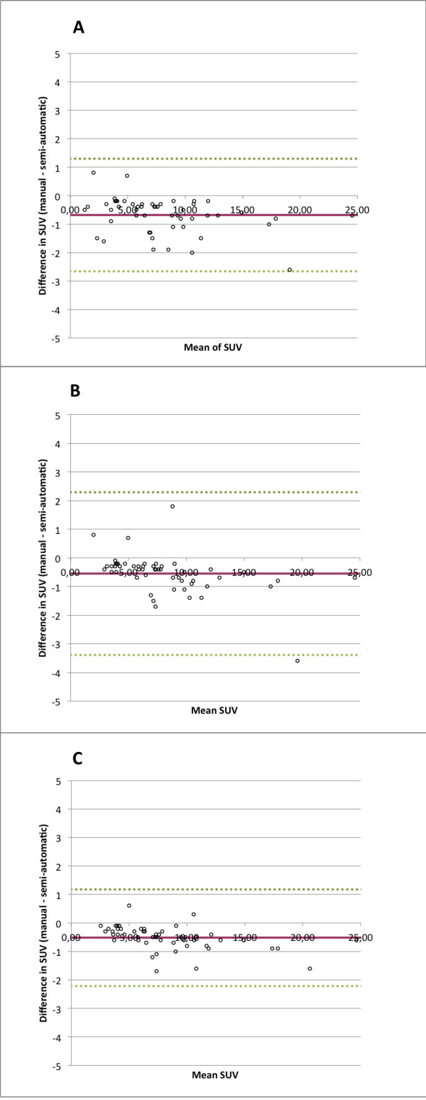
Background: Changes in maximum standardised uptake values (SUVmax) between serial PET/CT studies are used to determine disease progression or regression in oncologic patients. To measure these changes manually can be time consuming in a clinical routine. A semi-automatic method for calculation of SUVmax in serial PET/CT studies was developed and compared to a conventional manual method. The semi-automatic method first aligns the serial PET/CT studies based on the CT images. Thereafter, the reader selects an abnormal lesion in one of the PET studies. After this manual step, the program automatically detects the corresponding lesion in the other PET study, segments the two lesions and calculates the SUVmax in both studies as well as the difference between the SUVmax values. The results of the semi-automatic analysis were compared to that of a manual SUVmax analysis using a Philips PET/CT workstation. Three readers did the SUVmax readings in both methods. Sixteen patients with lung cancer or lymphoma who had undergone two PET/CT studies were included. There were a total of 26 lesions.
Results: Linear regression analysis of changes in SUVmax show that intercepts and slopes are close to the line of identity for all readers (reader 1: intercept = 1.02, R2 = 0.96; reader 2: intercept = 0.97, R2 = 0.98; reader 3: intercept = 0.99, R2 = 0.98). Manual and semi-automatic method agreed in all cases whether SUVmax had increased or decreased between the serial studies. The average time to measure SUVmax changes in two serial PET/CT examinations was four to five times longer for the manual method compared to the semi-automatic method for all readers (reader 1: 53.7 vs. 10.5 s; reader 2: 27.3 vs. 6.9 s; reader 3: 47.5 vs. 9.5 s; p < 0.001 for all).
Conclusions: Good agreement was shown in assessment of SUVmax changes between manual and semi-automatic method. The semi-automatic analysis was four to five times faster to perform than the manual analysis. These findings show the feasibility of using semi-automatic methods for calculation of SUVmax in clinical routine and encourage further development of programs using this type of methods.
Figures

Similar articles
-
Computer-assisted quantitative evaluation of therapeutic responses for lymphoma using serial PET/CT imaging.Acad Radiol. 2010 Apr;17(4):479-88. doi: 10.1016/j.acra.2009.10.026. Epub 2010 Jan 12. Acad Radiol. 2010. PMID: 20060747 Free PMC article. Clinical Trial.
-
Assessment of combination of contrast-enhanced magnetic resonance imaging and positron emission tomography/computed tomography for evaluation of ovarian masses.Invest Radiol. 2014 Aug;49(8):524-31. doi: 10.1097/RLI.0000000000000050. Invest Radiol. 2014. PMID: 24637584
-
Phantom validation of coregistration of PET and CT for image-guided radiotherapy.Med Phys. 2004 May;31(5):1083-92. doi: 10.1118/1.1688041. Med Phys. 2004. PMID: 15191296
-
Prognostic value of SUVmax and metabolic tumor volume on 18F-FDG PET/CT in early stage non-small cell lung cancer patients without LN metastasis.Biomed Mater Eng. 2014;24(6):3091-103. doi: 10.3233/BME-141131. Biomed Mater Eng. 2014. PMID: 25227018
-
Feasibility and performance of an adaptive contrast-oriented FDG PET/CT quantification technique for global disease assessment of malignant pleural mesothelioma and a brief review of the literature.Hell J Nucl Med. 2015 Jan-Apr;18(1):11-8. doi: 10.1967/s002449910162. Epub 2015 Feb 13. Hell J Nucl Med. 2015. PMID: 25679073 Review.
References
Publication types
MeSH terms
Substances
LinkOut - more resources
Full Text Sources
Medical
