. 2012 Apr 3:13:24.
doi: 10.1186/1471-2350-13-24.

Mutations in the potassium channel subunit KCNE1 are associated with early-onset familial atrial fibrillation

Affiliations

Mutations in the potassium channel subunit KCNE1 are associated with early-onset familial atrial fibrillation

Morten S Olesen et al. BMC Med Genet. .

Abstract

Background: Atrial fibrillation (AF) is the most common arrhythmia. The potassium current IKs is essential for cardiac repolarization. Gain-of-function mutations in KV7.1, the pore-forming α-subunit of the IKs channel, have been associated with AF. We hypothesized that early-onset lone AF is associated with mutations in the IKs channel regulatory subunit KCNE1.

Methods: In 209 unrelated early-onset lone AF patients (< 40 years) the entire coding sequence of KCNE1 was bidirectionally sequenced. We analyzed the identified KCNE1 mutants electrophysiologically in heterologous expression systems.

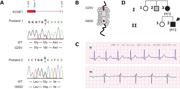
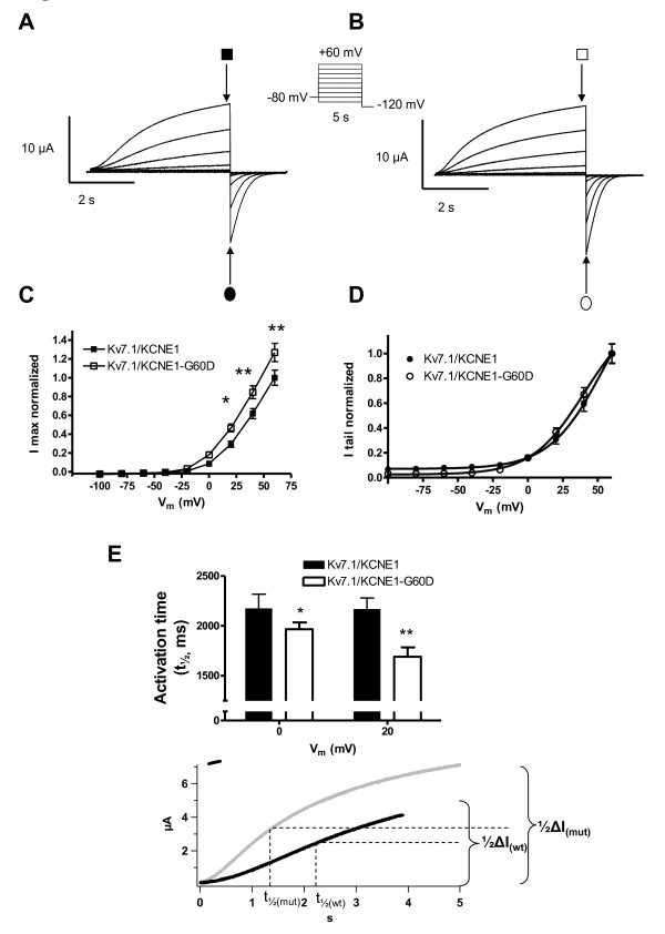
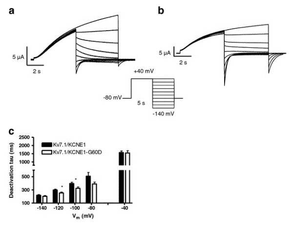
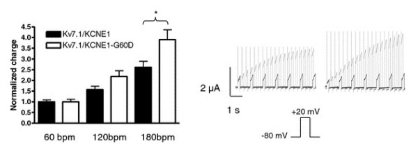
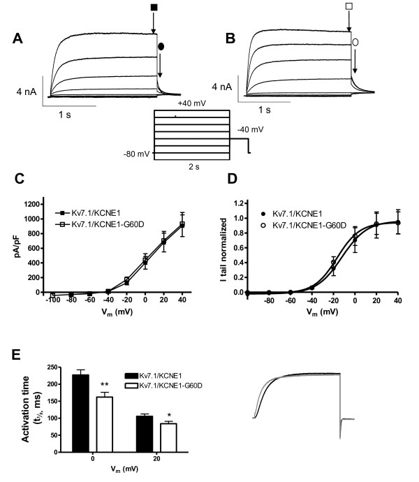
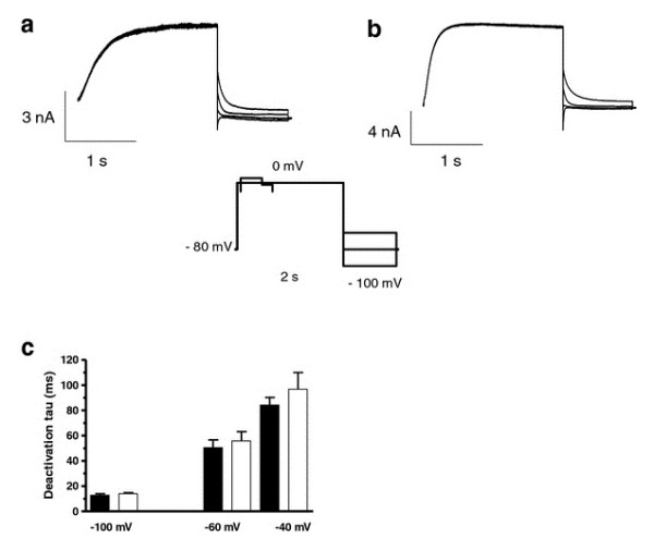
Results: Two non-synonymous mutations G25V and G60D were found in KCNE1 that were not present in the control group (n = 432 alleles) and that have not previously been reported in any publicly available databases or in the exom variant server holding exom data from more than 10.000 alleles. Proband 1 (female, age 45, G25V) had onset of paroxysmal AF at the age of 39 years. Proband 2 (G60D) was diagnosed with lone AF at the age of 33 years. The patient has inherited the mutation from his mother, who also has AF. Both probands had no mutations in genes previously associated with AF. In heterologous expression systems, both mutants showed significant gain-of-function for IKs both with respect to steady-state current levels, kinetic parameters, and heart rate-dependent modulation.

Conclusions: Mutations in KV7.1 leading to gain-of-function of IKs current have previously been described in lone AF, yet this is the first time a mutation in the beta-subunit KCNE1 is associated with the disease. This finding further supports the hypothesis that increased potassium current enhances AF susceptibility.


Figures

Figure 1
Figure 1
Clinical characterization and genetic analysis of probands. A: DNA sequence analysis. B: Positions of mutations indicated in schematic of protein topology. C: ECG from proband 2 (paper speed 25 mm/s, 1 mV/mm). D: Pedigree of the family with the novel KCNE1 G60D mutation. Squares: male, circles: female family members, respectively. Arrow indicates the proband 2. Solid black symbols indicate the presence of AF, open symbols: unaffected members, gray: AF history; (+/-): presence of the heterozygous mutation for persons with DNA samples available for testing.
Figure 2
Figure 2
Comparison of IKs-WT and IKs-G60D channel currents. Representative current traces for KCNE1-WT (A) and KCNE1-G60D (B) channel subunits co-expressed with KV7.1 in X.laevis oocytes in a 1:1 molar ratio. Current step protocol is shown as inset. C: I/V relationship for IKs-WT (n = 21) and IKs-G60D (n = 23) as determined from peak currents (open and filled squares). D: Voltage-dependence of IKs-WT and IKs-G60D channel activation as determined from tail currents (open and filled circles). E: Activation rise time, determined as the time to 1/2 max following a depolarization to 0 mV or +20 mV.
Figure 3
Figure 3
Comparison of IKs-WT and IKs-G60D channel deactivation. KCNE1-WT (A) and KCNE1-G60D (B) channel subunits co-expressed with KV7.1 in X.laevis oocytes in a 1:1 molar ratio. Current step protocol is shown as inset. (C) Enlargement of the tail-currents normalized to maximum current amplitude (gray: IKs-WT; black: IKs-G60D). Deactivation time constants (tau) were obtained by fitting the tail-current traces to a mono-exponential function (D).
Figure 4
Figure 4
Frequency dependence of conduction. Three different voltage protocols were applied (60 bpm (+20 mV for 200 ms, followed by 800 ms at -80 mV); 120 bpm (+20 mV for 180 ms, followed by 320 ms at -80 mV); 180 bpm (+20 mV for 150 ms, followed by 180 ms at -80 mV)). The bar graph summarizes the amount of charge conducted by KV7.1-WT/KCNE1 (black; n = 10) or KV7.1/KCNE1-G60D (white; n = 15) channels in the first 130 ms after the capacitive spike (10 ms) of the pulse at the 7th second when normalized to the charge carried at 60 bpm. Representative current traces recorded (left: KV7.1/KCNE1; right: KV7.1/KCNE1-G60D) using the 60 bpm (black) and 120 bpm (gray) protocols are shown on the right.
Figure 5
Figure 5
Comparison of IKs-WT and IKs-G60D channel currents at 37°C. KCNE1-WT (A) and KCNE1-G60D (B) channel subunits co-expressed with KV7.1 in CHO cells. Current protocol shown as inset. C: I/V relationship for IKs-WT (n = 15) and IKs-G60D (n = 12). Currents were measured at the end of the 2-second test pulse (open and filled squares) and normalized to cell size. D: Voltage-dependence of IKs-WT and IKs-G60D channel activation. Peak tail-currents were measured at -40 mV (open and filled circles), and the normalized data were fit to a two-state Boltzmann distribution. E: Activation rise time, determined as the time to 1/2 max following a depolarization to 0 or +20 mV.
Figure 6
Figure 6
Comparison of IKs-WT and IKs-G60D channel deactivation kinetics. KCNE1-WT (A) and KCNE1-G60D (B) channel subunits co-expressed with KV7.1 in CHO cells. Currents were elicited by clamping the cells for 2 s at 0 mV, followed by a 1 s step to test potentials ranging from -100 to -20 mV in 20 mV increments, at 36 ± 1°C. (C) Enlargement of the tail-currents normalized to maximum current amplitude (gray: IKs-WT; black: IKs-G60D). Deactivation time constants (tau) were obtained by fitting the tail-current traces to a mono-exponential function (D).

Similar articles

Cited by

References

    1. Benjamin EJ, Wolf PA, D'Agostino RB. et al.Impact of atrial fibrillation on the risk of death: the Framingham heart study. Circulation. 1998;98(10):946–952. - PubMed
    1. Fuster V, Rydén LE, Cannom DS. et al.ACC/AHA/ESC 2006 guidelines for the management of patients with atrial fibrillation-excutive summary. Rev Port Cardiol. 2007;26(4):383–446. - PubMed
    1. Stewart S, Hart CL, Hole DJ, McMurray JJ. Population prevalence, incidence, and predictors of atrial fibrillation in the Renfrew/Paisley study. Heart. 2001;86(5):516–521. doi: 10.1136/heart.86.5.516. - DOI - PMC - PubMed
    1. Go AS, Hylek EM, Phillips KA. et al.Prevalence of diagnosed atrial fibrillation in adults: national implications for rhythm management and stroke prevention: the anticoagulation and risk factors in atrial fibrillation (ATRIA) study. JAMA. 2001;285(18):2370–2375. doi: 10.1001/jama.285.18.2370. - DOI - PubMed
    1. Psaty BM, Manolio TA, Kuller LH. et al.Incidence of and risk factors for atrial fibrillation in older adults. Circulation. 1997;96(7):2455–2461. - PubMed

Publication types

MeSH terms