Review
. 2012 Apr 2;197(1):11-8.
doi: 10.1083/jcb.201108006.

Breaking the ties that bind: new advances in centrosome biology

Affiliations
Review

Breaking the ties that bind: new advances in centrosome biology

Balca R Mardin et al. J Cell Biol. .

Abstract

The centrosome, which consists of two centrioles and the surrounding pericentriolar material, is the primary microtubule-organizing center (MTOC) in animal cells. Like chromosomes, centrosomes duplicate once per cell cycle and defects that lead to abnormalities in the number of centrosomes result in genomic instability, a hallmark of most cancer cells. Increasing evidence suggests that the separation of the two centrioles (disengagement) is required for centrosome duplication. After centriole disengagement, a proteinaceous linker is established that still connects the two centrioles. In G2, this linker is resolved (centrosome separation), thereby allowing the centrosomes to separate and form the poles of the bipolar spindle. Recent work has identified new players that regulate these two processes and revealed unexpected mechanisms controlling the centrosome cycle.


Figures

Figure 1.
Figure 1.
The centrosome cycle of animal cells. Main events in the centrosome cycle are highlighted along with the key players that have been implicated in each process. Green-filled regions represent the centrin-positive distal regions of the centriole. As cells exit mitosis the daughter centriole disengages from the mother centriole, losing its orthogonal connection (centriole disengagement). Upon disengagement, the daughter centriole is connected to the mother by a flexible linker (pink strands). Centriole assembly factors accumulate in S phase and new centrioles are formed and gradually elongate throughout S and G2. At the G2/M transition the flexible linker that holds the centriole pairs together is lost (linker dissolution) and the centrioles accumulate more PCM (maturation) and constitute the poles of the mitotic spindle.
Figure 2.
Figure 2.
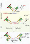
Centriole disengagement and duplication. Main players of centriole disengagement are depicted. During mitosis, Plk1 and separase consecutively disjoin mother and daughter centrioles. In addition, Plk1-mediated modification of the centrioles defines their capability of becoming a fully functional centrosome. In late mitosis the main centriole assembly factor hSas-6 is degraded and its levels are kept low by APC/C- and SCF-mediated proteolysis until the duplication can be initiated. In addition, activation of Plk-4 inhibits this negative regulation to allow cartwheel formation.
Figure 3.
Figure 3.
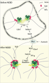
Centrosome separation before and after NEBD. Plk1 has a central role in regulating the centrosome separation via different pathways. In G2, Plk1 activates Nek9/6/7 cascade to regulate the accumulation of Eg5. Moreover, Cdk1 is important for Eg5 binding to MTs. After NEBD, Plk1 regulates the phospho-balance on the centrosomal linker by controlling the association of Nek2 with PP1γ through phosphorylation of Mst2. In addition, Plk1 is involved in the targeting of Eg5 to the spindle poles.

Similar articles

Cited by

References

    1. Alliegro M.C., Alliegro M.A., Palazzo R.E. 2006. Centrosome-associated RNA in surf clam oocytes. Proc. Natl. Acad. Sci. USA. 103:9034–9038 10.1073/pnas.0602859103 - DOI - PMC - PubMed
    1. Azimzadeh J., Bornens M. 2007. Structure and duplication of the centrosome. J. Cell Sci. 120:2139–2142 10.1242/jcs.005231 - DOI - PubMed
    1. Azimzadeh J., Marshall W.F. 2010. Building the centriole. Curr. Biol. 20:R816–R825 10.1016/j.cub.2010.08.010 - DOI - PMC - PubMed
    1. Bahe S., Stierhof Y.D., Wilkinson C.J., Leiss F., Nigg E.A. 2005. Rootletin forms centriole-associated filaments and functions in centrosome cohesion. J. Cell Biol. 171:27–33 10.1083/jcb.200504107 - DOI - PMC - PubMed
    1. Bahmanyar S., Kaplan D.D., Deluca J.G., Giddings T.H., Jr, O’Toole E.T., Winey M., Salmon E.D., Casey P.J., Nelson W.J., Barth A.I. 2008. beta-Catenin is a Nek2 substrate involved in centrosome separation. Genes Dev. 22:91–105 10.1101/gad.1596308 - DOI - PMC - PubMed

Publication types