. 2012 Apr 2;197(1):37-44.
doi: 10.1083/jcb.201112094.

Ovastacin, a cortical granule protease, cleaves ZP2 in the zona pellucida to prevent polyspermy

Affiliations

Ovastacin, a cortical granule protease, cleaves ZP2 in the zona pellucida to prevent polyspermy

Anna D Burkart et al. J Cell Biol. .

Abstract

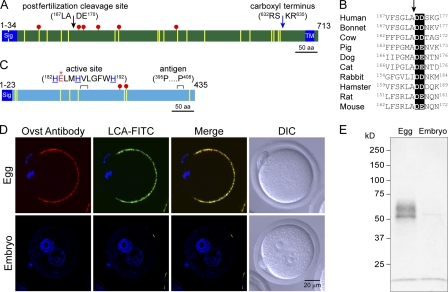
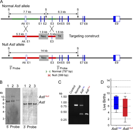
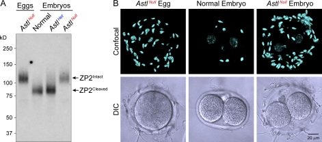
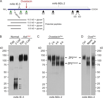
The mouse zona pellucida is composed of three glycoproteins (ZP1, ZP2, and ZP3), of which ZP2 is proteolytically cleaved after gamete fusion to prevent polyspermy. This cleavage is associated with exocytosis of cortical granules that are peripherally located subcellular organelles unique to ovulated eggs. Based on the cleavage site of ZP2, ovastacin was selected as a candidate protease. Encoded by the single-copy Astl gene, ovastacin is an oocyte-specific member of the astacin family of metalloendoproteases. Using specific antiserum, ovastacin was detected in cortical granules before, but not after, fertilization. Recombinant ovastacin cleaved ZP2 in native zonae pellucidae, documenting that ZP2 was a direct substrate of this metalloendoprotease. Female mice lacking ovastacin did not cleave ZP2 after fertilization, and mouse sperm bound as well to Astl-null two-cell embryos as they did to normal eggs. Ovastacin is a pioneer component of mouse cortical granules and plays a definitive role in the postfertilization block to sperm binding that ensures monospermic fertilization and successful development.


Figures

Figure 1.
Figure 1.
Localization of ovastacin. (A) Mouse ZP2 (713 aa) is proteolytically processed by cleavage of a signal peptide (Sig) and at a bibasic motif (blue arrow) upstream of a transmembrane (TM) domain to release the ectodomain (ZP235-633) that participates in the extracellular zona pellucida. After fertilization, ZP2 is cleaved upstream of a diacidic motif (169DE170). Conserved cysteine residues (yellow vertical lines) and potential N-linked glycosylation sites (red circles) are indicated. (B) The diacidic (shaded) proteolytic cleavage site (arrow) in the N-terminal region of mouse ZP2 is conserved in human, bonnet monkey, cow, pig, dog, cat, rabbit, hamster, and rat. (C) A schematic of mouse ovastacin including a signal peptide, a Zn+2-binding (blue histidine residues) enzyme-active site (red glutamic acid with an asterisk), and the antigen (395PLALFPEARDKPAP408) used to generate a rabbit pAb. Cysteine residues and potential glycosylation sites are indicated as in A. (D) Unfertilized eggs and one-cell embryos from normal mice were imaged by confocal microscopy and differential interference contrast (DIC) after staining with rabbit anti-ovastacin (Ovst) antibody, LCA-FITC (a marker of cortical granules), and the nuclear stain Hoechst 33342. (E) An immunoblot of lysates from unfertilized eggs (150) and two-cell embryos (150) was probed with antibody to ovastacin.
Figure 2.
Figure 2.
Establishment of AstlNull mice. (A) A schematic of the normal and AstlNull alleles after targeting with a construct containing positive (phosphoglucokinase [PGK]-Neo) and negative (MC1-TK) selectable markers. The thicker lines represent the 5.3- and 1.5-kbp homologous arms that are 5′ and 3′, respectively, to the Neo cassette. 5′ (407 bp; 5 bp outside of the targeting construct) and 3′ (368 bp; 97 bp outside of the targeting construct) probes were used for Southern blot hybridization. Arrows indicate SspI (S) restriction endonuclease sites. PCR genotyping was performed using primers for the normal allele (797 bp) and null allele (399 bp). Alt., alternative. (B) Southern blot hybridization of ES cell DNA from two successfully targeted clones (1 and 2) and one normal control (3) detected the normal allele as 7.7- and 6.3-kbp fragments with the 5′ and 3′ 32P-labeled probes, respectively. The AstlNull allele was detected as a 14-kbp fragment with either the 5′ or the 3′ probe. (C) PCR genotyping of mouse tail DNA detected the normal (797 bp) and null (399 bp) Astl alleles. (D) The size of litters from AstlHet and AstlNull female mice (five per group) mated with normal males over a period of 8–10 mo. Box plots reflect the median (line) and data points within the 10th and 90th percentiles (error bars). Boxes include the middle two quartiles, and outliers are indicated by dots.
Figure 3.
Figure 3.
Localization of ovastacin in normal and AstlNull mice. (A) Unfertilized eggs and two-cell embryos from normal, AstlHet, and AstlNull mice were collected and viewed by confocal and differential interference contrast (DIC) microscopy. Eggs and embryos were stained with rabbit anti-ovastacin (Ovst) antibody, LCA-FITC, and Hoechst 33342. Peripherally located cortical granules stained with LCA-FITC (white arrows) were observed in normal and AstlHet but not AstlNull eggs. The presence or absence of ovastacin correlated with the detection of LCA-FITC (black arrows in differential interference contrast/merge). (B) AstlHet and AstlNull eggs were stained with WGA lectin and imaged by EM. Cortical granules beneath the ooplasm (arrows) and the extracellular zona pellucida stained with WGA are shown. Mitochondria do not stain and serve as a negative control. After fertilization, cortical granules were absent from AstlHet two-cell embryos, although WGA continued to stain the zona pellucida. Bars, 0.5 µm. (C, top) Immunoblots of insect cell supernatant lacking (control [Ctrl]) or containing (Ovst) recombinant ovastacin probed with antibody to ovastacin (left) or with LCA lectin (right). (bottom) Immunoblots are the same after immunoprecipitation of normal ovarian lysates with antibody to ovastacin.
Figure 4.
Figure 4.
ZP2 cleavage and sperm binding to AstlNull two-cell embryos. (A) Immunoblot of lysates from eggs (20) and two-cell embryos (20) from normal, AstlHet, and AstlNull mice probed with an mAb specific for the C-terminal region of mouse ZP2 (M2c.2). Intact ZP2 is 120 kD, and the cleaved C-terminal fragment of ZP2 is 90 kD. (B) Eggs and two-cell embryos from AstlNull and two-cell embryos from normal mice were incubated (1 h) with capacitated sperm. After washing with a wide-bore pipette to remove all but two to six sperm on normal two-cell embryos (negative control), eggs and embryos were stained with Hoechst 33342, and bound sperm were presented as z projections of 5-µm confocal optical sections and differential interference contrast (DIC) images.
Figure 5.
Figure 5.
Cleavage of ZP2 by ovastacin. (A) A schematic of mouse ZP2 with eight diacidic motifs (triangles), the initial cleavage site of ovastacin, the binding sites of mAbs IE-3 and M2c.2 N terminal and C terminal to the initial cleavage site, and the localization of the single N-glycan in the N terminus. The potential size of peptides resulting from cleavage of the N-terminal fragment with ovastacin detected by IE-3 is shown on the bottom. (B) Immunoblot of eggs (15) or two-cell embryos (15) from normal and AstlNull females stained with IE-3 mAb that detects N-terminal fragments of ZP2. The top bands (120 kD) are intact ZP2, and bottom bands are a heterogeneous mixture of cleaved N-terminal fragments. Arrows indicate the four peptides detected on the immunoblot probed with IE-3. (C) Isolated zonae pellucidae (15–20) were incubated with partially purified recombinant ovastacin (OvastacinRec) at 37°C for 0, 2, 4, and 8 h. (D) Same as in C but with insect cell supernatant lacking (control [Ctrl]) or containing (OvstRec) recombinant ovastacin after 4 h of incubation with zonae pellucidae from normal (Norm) or AstlNull eggs. (C and D) Cleavage was detected by immunoblotting with M2c.2 to detect the ZP2 C-terminal fragment.

Similar articles

Cited by

References

    1. Baibakov B., Gauthier L., Talbot P., Rankin T.L., Dean J. 2007. Sperm binding to the zona pellucida is not sufficient to induce acrosome exocytosis. Development. 134:933–943 10.1242/dev.02752 - DOI - PubMed
    1. Barros C., Yanagimachi R. 1971. Induction of zona reaction in golden hamster eggs by cortical granule material. Nature. 233:268–269 10.1038/233268a0 - DOI - PubMed
    1. Bauskin A.R., Franken D.R., Eberspaecher U., Donner P. 1999. Characterization of human zona pellucida glycoproteins. Mol. Hum. Reprod. 5:534–540 10.1093/molehr/5.6.534 - DOI - PubMed
    1. Bleil J.D., Wassarman P.M. 1980. Structure and function of the zona pellucida: Identification and characterization of the proteins of the mouse oocyte’s zona pellucida. Dev. Biol. 76:185–202 10.1016/0012-1606(80)90371-1 - DOI - PubMed
    1. Bleil J.D., Beall C.F., Wassarman P.M. 1981. Mammalian sperm-egg interaction: Fertilization of mouse eggs triggers modification of the major zona pellucida glycoprotein, ZP2. Dev. Biol. 86:189–197 10.1016/0012-1606(81)90329-8 - DOI - PubMed

Publication types