. 2012 Apr 2;197(1):45-57.
doi: 10.1083/jcb.201112145.

Nuclear translation visualized by ribosome-bound nascent chain puromycylation

Affiliations

Nuclear translation visualized by ribosome-bound nascent chain puromycylation

Alexandre David et al. J Cell Biol. .

Abstract

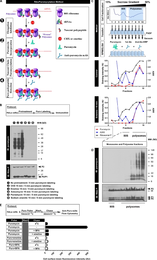
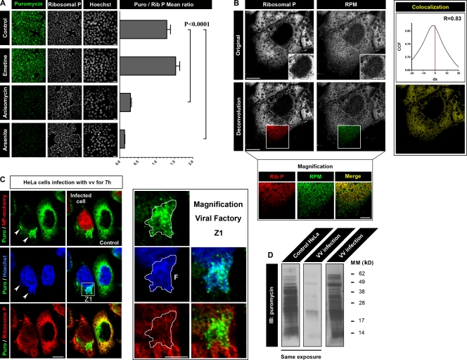
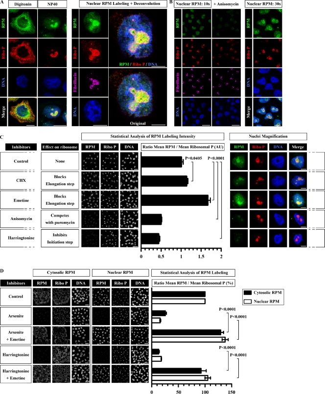
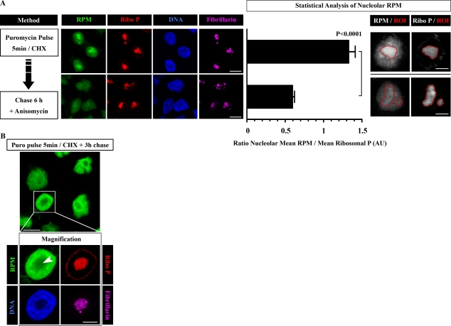
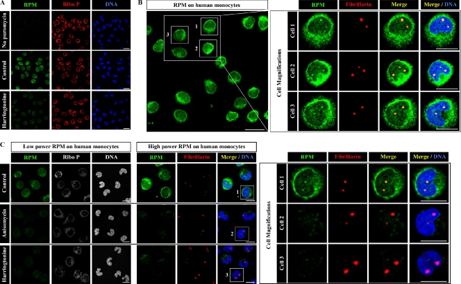
Whether protein translation occurs in the nucleus is contentious. To address this question, we developed the ribopuromycylation method (RPM), which visualizes translation in cells via standard immunofluorescence microscopy. The RPM is based on ribosome-catalyzed puromycylation of nascent chains immobilized on ribosomes by antibiotic chain elongation inhibitors followed by detection of puromycylated ribosome-bound nascent chains with a puromycin (PMY)-specific monoclonal antibody in fixed and permeabilized cells. The RPM correlates localized translation with myriad processes in cells and can be applied to any cell whose translation is sensitive to PMY. In this paper, we use the RPM to provide evidence for translation in the nucleoplasm and nucleolus, which is regulated by infectious and chemical stress.


Figures

Figure 1.
Figure 1.
Characterizing the RPM biochemically. (A) Schematic representation of the RPM. After freezing polysomes with an elongation inhibitor (step 1), PMY is added (step 2) to living cells or subcellular fractions, and nascent chains are puromycylated through ribosome catalysis (step 3). The anti-PMY mAb 12D10 (or other PMY mAbs that we have generated) detects puromycylated nascent chains via immunoblotting or indirect immunofluorescence (step 4). (B) Anti-PMY immunoblotting (IB) of total HeLa cell lysates from cells incubated with PMY for 5 min and other inhibitors as indicated. The smear of proteins represents C-terminally puromycylated nascent chains released from ribosomes. Chain elongation inhibitors do not effect (CHX) or enhance (emetine) nascent chain puromycylation, whereas protein synthesis inhibitors that deplete nascent chains by blocking initiation while allowing chain elongation and completion (pactamycin, direct initiation inhibitor, and arsenite, indirect initiation inhibitor) prevent puromycylation. Anisomycin blocks puromycylation by competing with PMY binding to ribosomes. On the bottom, blotting with anti–ribosomal P (Ribo P) human autoreactive antisera shows that the results cannot be attributed to lane loading discrepancies. (C) HeLa cells incubated or not incubated with emetine for 15 min were lysed and fractionated on 15–50% sucrose gradients. Fractions were bound to PVDF 96-well plates and incubated with PMY, which results in ribosome-catalyzed nascent chain puromycylation. Ribosomes were detected by A260 of fractions or by ELISA for the ribosomal P proteins (here resolved into the three known species) as detected by human autoimmune antibodies, which establishes that monosomes and 60S subunits bind well to PVDF. Puromycylation was detected by ELISA for PMY using 12D10 and clearly demonstrates that monosomes and free 60S subunits do not stably associate with PMY, which requires nascent chain puromycylation. (D) HeLa cells incubated with emetine were lysed and fractionated on 15–50% sucrose gradients. Monosome- and polysome-containing fractions were labeled with PMY on ice, and nascent chains were identified by immunoblotting with 12D10. Only polysomes demonstrate a significant anti-PMY signal, and from the pattern, it is clear that binding is based on nascent chain puromycylation. As expected, the mean size of puromycylated proteins increases with polysome size because, on average, faster sedimenting polysomes possess more ribosomes, translating longer mRNAs. (E) HeLa cells were pulsed with PMY with or without emetine or BFA. Cells were washed and chased with or without emetine/BFA. The control sample (pulse only, labeled 4°C) was kept cold during the chase. Expression of PMY (Puro) on the surface of live cells was determined by flow cytometry using 12D10. In the absence of inhibitors, some puromycylated nascent chains are sufficiently native to be delivered to and expressed on the cell surface with their C-terminal PMY exposed for detection (the basis for the SUnSET assay). Emetine blocks surface expression to the same extent as low temperature or BFA, which completely blocks egress of membrane proteins from the ER. This experiment functionally establishes that emetine prevents release of puromycylated nascent chains from ribosomes, as determined by the proxy population of cell surface proteins detected by the SUnSET method. Error bars represent the standard deviation of triplicate samples. Ab, antibody; AU, arbitrary units; MM, molecular mass.
Figure 2.
Figure 2.
Live-cell RPM detects translating ribosomes. (A) HeLa cells pretreated for 15 min with the inhibitor indicated were incubated with PMY (Puro) for 5 min at 37°C before digitonin extraction and PFA fixation. Samples were then incubated with 12D10 to detect PMY. For each condition, multiple fields were acquired, and the mean fluorescence ratio of PMY/ribosome staining for each field was quantitated using ImageJ. Values are plotted on the right (means ± SEM). Statistical analysis, two-tailed unpaired t test. Bars, 20 µm (B) Higher magnification images of a HeLa cell pretreated with emetine and labeled live with PMY at 37°C as in A. (bottom) The same image after deconvolution to minimize unfocused light from other focal planes. Nuclear magnification in top insets show weak but well above background RPM and ribosomal P (Rib P) labeling. Although overall colocalization is extensive (right column), magnification shows that there are imbalances in intensity of the staining. Bars: (main images and top insets) 10 µm; (bottom magnification) 5 µm. (C) Active translation was localized in HeLa cells 7 h after infection with a recombinant VV (rVV) expressing NP-mCherry by live RPM staining. In the images shown, only one of the two cells is infected, as demonstrated by the presence of NP-mCherry in the nucleus. Translation is nearly completely localized to viral factories in infected cells, identified as juxtanuclear DNA-positive structures (arrowheads). Even within a factory, ribosomes display different RPM intensities, indicative of differential translation rates and seen in the zoomed image, Z1. Bars: (main images) 10 µm; (Z1 images) 2 µm. F, factory. (D) Uninfected or VV-infected HeLa cells were labeled with PMY after pretreatment with emetine and were extracted directly in culture flasks with 1% NP-40. Puromycylated nascent chains were analyzed by immunoblotting with 12D10. VV infection diminishes the total PMY signal consistent with its effect on overall translation, and the translation profile differs from uninfected cells, consistent with detection of viral versus host proteins, as clearly shown from a longer exposure (right) to normalize levels of puromycylation. CCF, cross-correlation function; IB, immunoblot; MM, molecular mass.
Figure 3.
Figure 3.
RPM detects nuclear/nucleolar translation. (A) HeLa cells were labeled live with emetine and PMY and processed for RPM staining using the regular digitonin extraction method or NP-40 extraction before fixation. (right) Higher magnification series of the nucleus stained after NP-40 extraction demonstrates intense RPM staining of a fibrillarin-positive nucleolus. Merged image is shown with and without deconvolution to minimize unfocused light. Bars: (main images) 10 µm; (large images) 5 µm. (B) HeLa cells were labeled with PMY for 10 or 30 s before NP-40 extraction. Pretreating cells with anisomycin, a competitive inhibitor of PMY, reduces RPM to background values, demonstrating the ribosome dependence of nuclear RPM staining. Bars: (left) 20 µm; (right) 10 µm. (C) HeLa cells pretreated for 20 min with the inhibitor indicated were incubated for 5 min with PMY before RPM staining using NP-40 extraction. Five fields were acquired for each condition, and the mean fluorescence ratio of PMY/ribosome staining for each field was quantitated using ImageJ software. Values are plotted on the right (means ± SEM). Statistical analysis, two-tailed unpaired t test. Bars: (left) 20 µm; (right) 5 µm. (D) HeLa cells pretreated for 20 min with the inhibitors indicated were incubated for 5 min with PMY before RPM staining using either digitonin or NP-40 extraction. Emetine blocks the inhibitory effects of arsenite or harringtonine on cytoplasmic and nuclear RPM staining (digitonin vs. NP-40 extraction), demonstrating that RPM staining is based on the presence of ribosome-associated nascent chains. As for C, five fields were acquired for each condition, and the mean fluorescence ratio of PMY/ribosome staining for each field was quantitated using ImageJ. Values are plotted on the right (means ± SEM), considering the control ratio as 100%. Statistical analysis, two-tailed unpaired t test. Bars, 20 µm. AU, arbitrary unit; Ribo P, ribosomal P.
Figure 4.
Figure 4.
Stress-regulated nuclear translation detected by RPM. (A) Uninfected HeLa cells or HeLa cells infected for 2, 4, or 5 h with rVV expressing NP-mCherry at a high MOI to infect all cells in fields before digitonin (cytoplasmic)- or NP-40 (nuclear)–based RPM staining. It is clear that nuclear RPM staining decreases in parallel with shunting of protein synthesis to viral factories and increased NP-mCherry expression. (B) HeLa cells were infected with rVV expressing NP-mCherry at a lower MOI to infect ∼50% of cells before nuclear RPM staining (NP-40 extraction). (left) At 6 h after infection, the three infected cells of the six in the field demonstrate greatly reduced RPM staining in the nucleoplasm and, particularly, nucleoli. At 7 h after infection, one infected cell (identified by its DNA labeled viral factory) of two in the field demonstrates no detectable nuclear RPM staining. Arrowheads show the nucleus of VV-infected cells as identified by DNA staining of factory. (C) Uninfected HeLa cells or HeLa cells infected for 7 h with recombinant VSV expressing NP-EGFP before RPM staining and extraction with either digitonin (cytoplasmic RPM) or NP-40 (nuclear RPM). Five fields were acquired for each condition, and the mean fluorescence ratio of PMY/ribosome staining for each field was quantitated using ImageJ. Values are plotted on the right (means ± SEM). Statistical analysis, two-tailed unpaired t test. VSV infection clearly results in diminished nuclear translation despite robust cytoplasmic synthesis of viral proteins, further ruling out the possibility that the nuclear/nucleolar RPM is caused by trafficking/trapping of cytoplasmic puromycylated proteins. (D) Uninfected HeLa cells or HeLa cells infected for 7 h with recombinant SFV expressing IAV NP before RPM staining and extraction with either digitonin (cytoplasmic RPM) or NP-40 (nuclear RPM). As previously reported (Berglund et al., 2007), when expressed by SFV, NP principally localizes to mitochondria because of downstream initiation (cytoplasmic staining), with a minor full-length cohort localizing to the nucleus (nuclear staining). In SFV-infected cells (arrowheads), it is clear that both cytoplasmic and nuclear RPM staining are greatly diminished compared with surrounding uninfected cells. High magnification of cytoplasmic RPM staining reveals multiple translation foci in which viral proteins are likely translated. (E) HeLa cells either pretreated for 15 min with the inhibitor indicated or infected with IAV (Flu) were incubated with PMY for 5 min at 37°C in the presence of emetine. In this experiment, cells were simultaneously fixed and permeabilized by incubation with PBS containing NP-40 and PFA. This allows antibody access to nucleoli and fixes cytoplasmic ribosomes to enable direct comparison between nuclear and cytoplasmic RPMs. Cells were stained for PMY (green), large ribosomal subunit (red), DNA, and fibrillarin. This experiment shows that the nucleolar RPM staining is robust compared with the cytoplasmic RPM and is inhibited in parallel with inhibitors that block protein synthesis. Diminished nucleolar RPM in IAV-infected cells provides additional evidence against trapping of cytoplasmic puromycylated proteins in the nucleoli during fixation. Bars, 10 µm. AU, arbitrary units; Ribo P, ribosomal P.
Figure 5.
Figure 5.
Nucleolar RPM labeling can be chased. (A) HeLa cells were pulse labeled for 5 min with PMY in the presence of CHX and then chased for 6 h in the presence of anisomycin to prevent further PMY incorporation. For time 0 and time 6 h, cells were RPM stained using NP-40 extraction. Nucleolar RPM staining was measured by defining regions of interest (ROIs) around 15 different nucleoli and calculating the ratio between RPM and ribosomal P (Ribo P) nucleolar mean intensities (mean ± SEM). Statistical analyses, two-tailed unpaired t test. (B) HeLa cells were labeled with PMY CHX for 5 min and then chased for 3 h in the absence of inhibitors. Cells were RPM stained using NP-40 extraction. The arrowhead shows the nucleolus. Bars: (A [left] and B [main image]) 10 µm; (A [right] and B [magnification]) 5 µm. AU, arbitrary unit.
Figure 6.
Figure 6.
Nuclear RPM staining in monocytes. (A) Elutriated human peripheral blood monocytes pretreated or not pretreated with harringtonine 15 min before RPM staining (5-min PMY pulse in the presence of emetine) were fixed and extracted simultaneously with polysome buffer containing PFA and digitonin. Note that in the absence of PMY no RPM staining is visible. (B) Same PMY staining protocol as in A. Intense RPM staining clearly colocalizes with nucleoli (red), which are smaller in monocytes than HeLa cells as shown by fibrillarin staining. (C) Elutriated monocytes pretreated or not pretreated with harringtonine or anisomycin for 15 min before RPM staining were fixed and extracted simultaneously with polysome buffer containing PFA and digitonin. RPM staining in cytoplasm and nucleoli is blocked by either inhibitor as seen in increasing magnifications from left to right, demonstrating the dependence of ribosome-catalyzed puromycylation of the nascent chain. Bars: (A and B [main image]) 20 µm; (B [magnification] and C) 10 µm. Ribo P, ribosomal P.

Comment in

Similar articles

Cited by

References

    1. Allfrey V.G. 1954. Amino acid incorporation by isolated thymus nuclei. I. The role of desoxyribonucleic acid in protein synthesis. Proc. Natl. Acad. Sci. USA. 40:881–885 10.1073/pnas.40.10.881 - DOI - PMC - PubMed
    1. Allfrey V.G., Mirsky A.E., Osawa S. 1955. Protein synthesis in isolated cell nuclei. Nature. 176:1042–1049 10.1038/1761042a0 - DOI - PubMed
    1. Apcher S., Daskalogianni C., Lejeune F., Manoury B., Imhoos G., Heslop L., Fåhraeus R. 2011. Major source of antigenic peptides for the MHC class I pathway is produced during the pioneer round of mRNA translation. Proc. Natl. Acad. Sci. USA. 108:11572–11577 10.1073/pnas.1104104108 - DOI - PMC - PubMed
    1. Berglund P., Finzi D., Bennink J.R., Yewdell J.W. 2007. Viral alteration of cellular translational machinery increases defective ribosomal products. J. Virol. 81:7220–7229 10.1128/JVI.00137-07 - DOI - PMC - PubMed
    1. Bhalla A.D., Gudikote J.P., Wang J., Chan W.K., Chang Y.F., Olivas O.R., Wilkinson M.F. 2009. Nonsense codons trigger an RNA partitioning shift. J. Biol. Chem. 284:4062–4072 10.1074/jbc.M805193200 - DOI - PMC - PubMed

Publication types