. 2012 Apr 2:8:42.
doi: 10.1186/1746-6148-8-42.

Transmissibility of caprine scrapie in ovine transgenic mice

Affiliations

Transmissibility of caprine scrapie in ovine transgenic mice

Katherine I O'Rourke et al. BMC Vet Res. .

Abstract

Background: The United States control program for classical ovine scrapie is based in part on the finding that infection is typically spread through exposure to shed placentas from infected ewes. Transmission from goats to sheep is less well described. A suitable rodent model for examining the effect of caprine scrapie isolates in the ovine host will be useful in the ovine scrapie eradication effort. In this study, we describe the incubation time, brain lesion profile, glycoform pattern and PrPSc distribution patterns in a well characterized transgenic mouse line (Tg338) expressing the ovine VRQ prion allele, following inoculation with brain from scrapie infected goats.

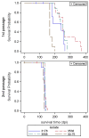
Results: First passage incubation times of caprine tissue in Tg338 ovinized mice varied widely but second passage intervals were shorter and consistent. Vacuolation profiles, glycoform patterns and paraffin-embedded tissue blots from terminally ill second passage mice derived from sheep or goat inocula were similar. Proteinase K digestion products of murine tissue were slightly smaller than the original ruminant inocula, a finding consistent with passage of several ovine strains in previous reports.

Conclusions: These findings demonstrate that Tg338 mice propagate prions of caprine origin and provide a suitable baseline for examination of samples identified in the expanded US caprine scrapie surveillance program.


Figures

Figure 1
Figure 1
Relative levels of PrPSc in brain homogenate from donor animals. Serial two-fold dilutions of brain homogenate from goat 3538 (A), 3558 (B), 30–75 (C) and reference sheep 3178 (D) were analyzed by western blot analysis and ELISA. Absorbance (A450) results from ELISA (−·-) are shown on the left y axis and density of the diglycosylated band determined from scans of western blot (vertical bars and inset image) on the right y axis. Starting wet weight equivalents per ELISA well or Western blot lane are shown on the x axis. Cut off limit for the ELISA (−−----------) was determined from the manufacturer’s negative control sample.
Figure 2
Figure 2
Kaplan-Meier survival curves for Tg338 mice inoculated with different isolates of small ruminant scrapie. Shown are the survival curves for first (upper panel) and second (lower panel) intracerebral passage in Tg338 mice including censored data. Scrapie isolates were derived from the brain of three clinical goats (broken lines: 3538, 3558 and 30–75) and a reference sheep (solid blue line: 3178). Note the reductions in survival times per isolate (days post-inoculation, dpi) as well as the reduction in survival time variation between isolates upon second passage. The color line code for each isolate source is indicated in the key.
Figure 3
Figure 3
Western blot analysis of brain from small ruminants and P2 Tg338 mice bioassay recipients. Western blot analysis of brain from goat 3538 (lane 1, 150 μg starting wet weight brain ), goat 3558 (lane 3, 250 μg ), goat 30–75 (lane 5, 250 μg) and sheep 3178 (lane 7, 350 μg), and brain from P2 mice from each of those 4 inoculum groups (lanes 2, 4 , 6 and 8, respectively, loaded with 60–100 μg brain) after digestion with proteinase K (50 μg/ml final concentration for goats and sheep, 100 μg/ml final concentration for mice) and detection with mAb F99/97.6.1, binding a carboxyl epitope (A) or mAb P4, binding an amino terminal epitope (B). Lane 9 contains brain (300 μg) from an age-matched uninoculated Tg338 mouse, homogenized and treated with proteinase K as described for tissue from P1 and P2 mice. Molecular mass markers are shown on the left.
Figure 4
Figure 4
Co-migration of brain homogenates from Tg338 mice inoculated by the intracerebral route or the intraperitoneal route. Brain homogenate from sheep 3178 (lanes 1, 3, 5 and 7, 350 μg starting wet weight per lane) and Tg338 mice (100 μg starting wet weight per lane) inoculated by the intracerebral route and assayed at passage 2 (lane 2) or passage 1 (lane 4). The same homogenate was inoculated intraperitoneally in Tg338 mice; first passage brain (lane 6) and spleen (lane 8) homogenates were assayed. Lane 9, brain homogenate (200 μg starting wet weight) from uninoculated Tg338 mouse. All tissues were digested with proteinase K (100 μg /ml final concentration for murine tissues and 50 μg/ml final concentration for ovine tissues). Filter was probed with mAb F99/97.6.1. Molecular mass markers are shown on the left.
Figure 5
Figure 5
Glycoform analysis of PrPSc in Tg338 mice inoculated with different isolates of small ruminant scrapie. Relative densities of unglycosylated, monoglycosylated and diglycosylated forms of PrPSc bands were obtained for each P2 mouse brain using an image analyzer. Green circle – inoculum group 3558; red circle – inoculum group 3538; black circle – inoculum group 30–75; blue circle – inoculum group 3178.
Figure 6
Figure 6
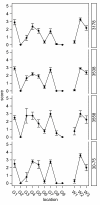
Histological vacuole scores by brain region for small ruminant scrapie in P2 Tg338 mice. Shown are the mean vacuolization scores with standard errors by Tg338 mouse brain region for scrapie isolates derived from the brain of three clinical goats and a reference sheep. Each isolate source case number is indicated to the right in each panel. Note the similar regional pattern of grey (G) and white (W) matter vacuole scores induced by goat and sheep-derived scrapie. G1, dorsal medulla nuclei; G2, cerebellar cortex of the folia including the granular layer adjacent to the fourth ventricle; G3, cortex of the superior colliculus; G4, hypothalamus; G5, thalamus; G6, hippocampus; G7, septal nuclei of the paraterminal body; G8, cerebral cortex (at the level of G4 and G5); G9, cerebral cortex (at the level of G7); W1, cerebellar white matter, W2, tegmentum; and W3, pyramidal tract.
Figure 7
Figure 7
Distribution of PK-resistant PrP labeling of PET blots. Shown is the neuroanatomic distribution of PrPSc at 4 different brain levels of P2 Tg338 mice representing inoculum groups 30–75 (A) and 3178 (B), and an age-matched uninoculated control group mouse (C). Brain sections were dissected at the levels of frontal cortex (level 1), hypothalamus (level 2), hippocampus (level 3) and cerebellum-medulla (level 4). Blots were probed with mAb F99/97.6.1. VP, ventral pallidum; HT, hypothalamus; MHN, medial hypothalamic nucleus; HN, medial habenular nucleus; NM, nucleus mammillaris; NR, nucleus rubor; SN, substantia nigra; NCG, nucleus corporis geniculati; DCN, deep cerebellar nuclei; M, medulla.

Similar articles

Cited by

References

    1. Will RG, Ironside JW, Zeidler M, Cousens SN, Estibeiro K, Alperovitch A, Poser S, Pocchiari M, Hofman A, Smith PG. A new variant of Creutzfeldt-Jakob disease in the UK. Lancet. 1996;347:921–925. doi: 10.1016/S0140-6736(96)91412-9. - DOI - PubMed
    1. Bruce ME, Will RG, Ironside JW, McConnell I, Drummond D, Suttle A, McCardie L, Chree A, Hope J, Birkett C. et al.Transmissions to mice indicate that ‘new variant’ CJD is caused by the BSE agent. Nature. 1997;389:498–501. doi: 10.1038/39057. - DOI - PubMed
    1. Peden AH, Head MW, Ritchie DL, Bell JE, Ironside JW. Preclinical vCJD after blood transfusion in a PRNP codon 129 heterozygous patient. Lancet. 2004;364:527–529. doi: 10.1016/S0140-6736(04)16811-6. - DOI - PubMed
    1. Oesch B, Westaway D, Walchi M, McKinley MP, Kent SBH, Aebersold R, Barry RA, Tempst P, Teplow DB, Hood LE. et al.A cellular gene encodes scrapie PrP27-30 protein. Cell. 1985;40:735–746. doi: 10.1016/0092-8674(85)90333-2. - DOI - PubMed
    1. Prusiner SB. Novel proteinaceous infectious particles cause scrapie. Science. 1982;216:136–144. doi: 10.1126/science.6801762. - DOI - PubMed

Publication types

LinkOut - more resources