DNA methylation profiling identifies global methylation differences and markers of adrenocortical tumors
- PMID: 22472567
- PMCID: PMC3387424
- DOI: 10.1210/jc.2011-3298
DNA methylation profiling identifies global methylation differences and markers of adrenocortical tumors
Abstract
Context: It is not known whether there are any DNA methylation alterations in adrenocortical tumors.
Objective: The objective of the study was to determine the methylation profile of normal adrenal cortex and benign and malignant adrenocortical tumors.
Methods: Genome-wide methylation status of CpG regions were determined in normal (n = 19), benign (n = 48), primary malignant (n = 8), and metastatic malignant (n = 12) adrenocortical tissue samples. An integrated analysis of genome-wide methylation and mRNA expression in benign vs. malignant adrenocortical tissue samples was also performed.
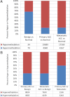
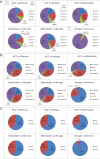
Results: Methylation profiling revealed the following: 1) that methylation patterns were distinctly different and could distinguish normal, benign, primary malignant, and metastatic tissue samples; 2) that malignant samples have global hypomethylation; and 3) that the methylation of CpG regions are different in benign adrenocortical tumors by functional status. Normal compared with benign samples had the least amount of methylation differences, whereas normal compared with primary and metastatic adrenocortical carcinoma samples had the greatest variability in methylation (adjusted P ≤ 0.01). Of 215 down-regulated genes (≥2-fold, adjusted P ≤ 0.05) in malignant primary adrenocortical tumor samples, 52 of these genes were also hypermethylated.
Conclusions: Malignant adrenocortical tumors are globally hypomethylated as compared with normal and benign tumors. Methylation profile differences may accurately distinguish between primary benign and malignant adrenocortical tumors. Several differentially methylated sites are associated with genes known to be dysregulated in malignant adrenocortical tumors.
Figures


Comment in
-
Adrenal gland: DNA methylation profiling--a new tool for adrenal tumour diagnosis?Nat Rev Endocrinol. 2012 Apr 24;8(6):320. doi: 10.1038/nrendo.2012.61. Nat Rev Endocrinol. 2012. PMID: 22525728 No abstract available.
References
-
- Androulakis II, Kaltsas GA, Markou A, Tseniklidi E, Kafritsa P, Pappa T, Papanastasiou L, Piaditis GP. 2011. The functional status of incidentally discovered bilateral adrenal lesions. Clin Endocrinol (Oxf) 75:44–49 - PubMed
-
- Grumbach MM, Biller BM, Braunstein GD, Campbell KK, Carney JA, Godley PA, Harris EL, Lee JK, Oertel YC, Posner MC, Schlechte JA, Wieand HS. 2003. Management of the clinically inapparent adrenal mass (“incidentaloma”). Ann Intern Med 138:424–429 - PubMed
-
- Icard P, Goudet P, Charpenay C, Andreassian B, Carnaille B, Chapuis Y, Cougard P, Henry JF, Proye C. 2001. Adrenocortical carcinomas: surgical trends and results of a 253-patient series from the French Association of Endocrine Surgeons Study Group. World J Surg 25:891–897 - PubMed
-
- Kloos RT, Gross MD, Francis IR, Korobkin M, Shapiro B. 1995. Incidentally discovered adrenal masses. Endocr Rev 16:460–484 - PubMed
-
- Assie G, Guillaud-Bataille M, Ragazzon B, Bertagna X, Bertherat J, Clauser E. 2010. The pathophysiology, diagnosis and prognosis of adrenocortical tumors revisited by transcriptome analyses. Trends Endocrinol Metab 21:325–334 - PubMed

