

Effects of donor characteristics and ex vivo expansion on canine mesenchymal stem cell properties: implications for MSC-based therapies
- PMID: 22472645
- PMCID: PMC3840229
- DOI: 10.3727/096368912X636821
Effects of donor characteristics and ex vivo expansion on canine mesenchymal stem cell properties: implications for MSC-based therapies
Abstract
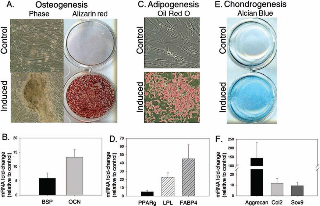
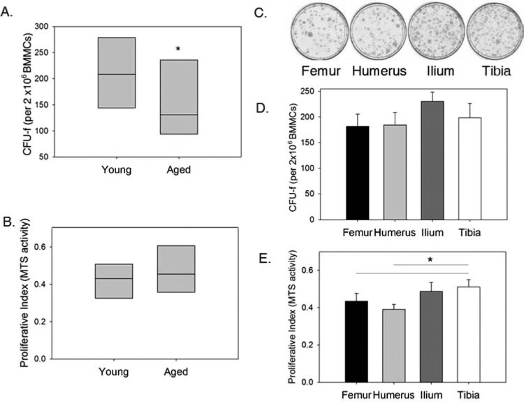
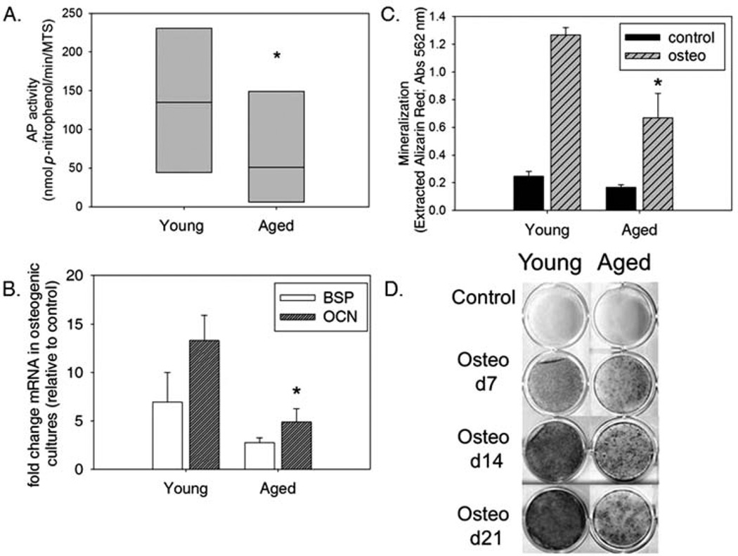
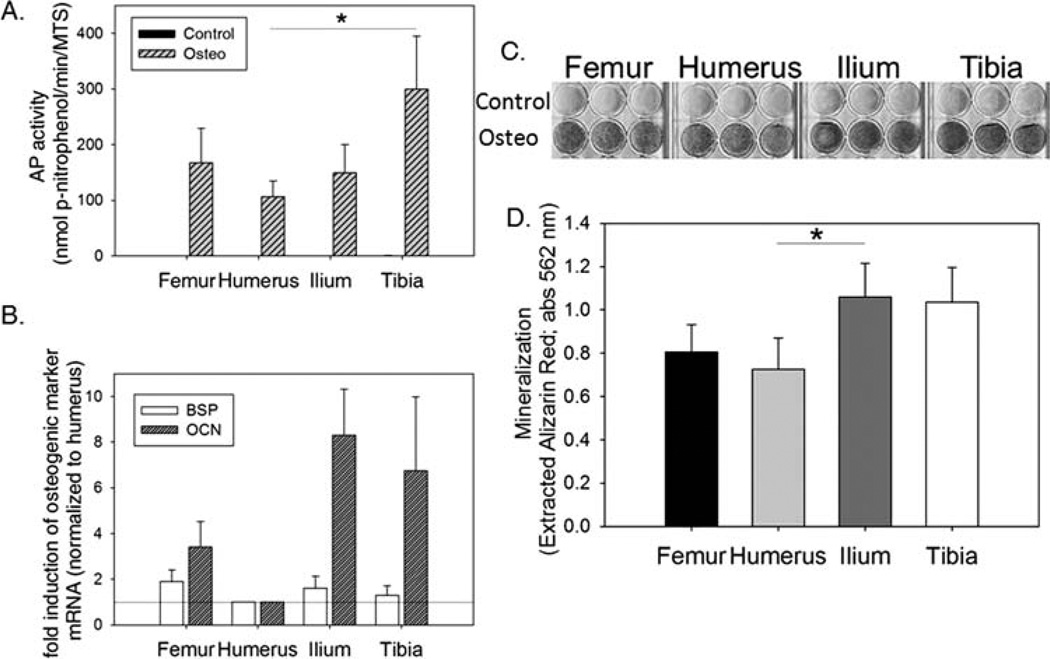
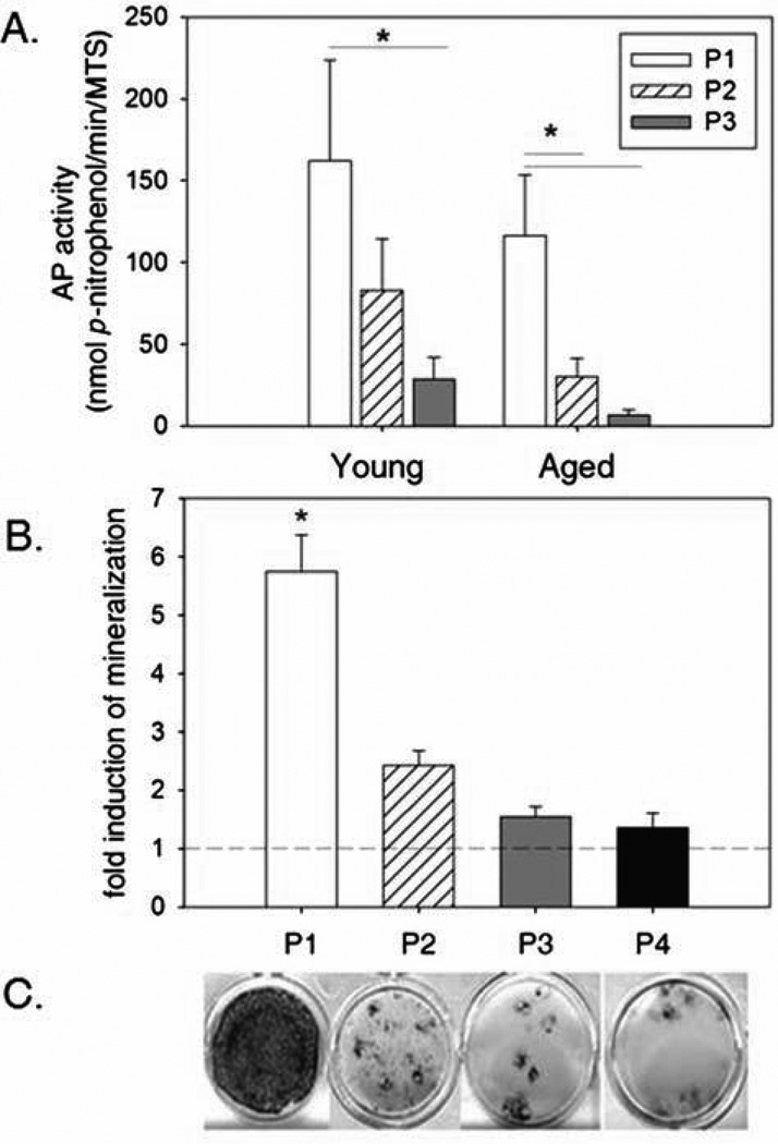
Clinical trials utilizing bone marrow-derived mesenchymal stem cell (BM-MSC) therapies show promise for treating a variety of pathologic conditions. Paramount to optimization of such cell-based therapies is a thorough understanding of MSC biology. Despite the tremendous potential that exists for the clinical use of canine BM-MSCs in veterinary medicine, as well as in preclinical studies for human medicine, relatively little information exists regarding basic biological properties of the cells. In this study, we compared the importance of donor characteristics (age and harvest site) and ex vivo expansion on canine BM-MSC frequency (CFU-f) and differentiation potential. Advancing age was found to have a negative effect on CFU-f as well as osteogenic potential. Site of harvest was also found to have significant effects on MSC properties. MSCs obtained from the humerus were found at the lowest frequency and were least osteogenic compared to those harvested from the tibia, femur, and ilium. Osteogenic potential diminished significantly by the third passage. These results suggest important donor parameters and culture effects to consider in translational studies examining MSC-based regenerative medical strategies.
Conflict of interest statement
The authors declare no conflicts of interest.
Figures
Similar articles
-
Instant stem cell therapy: characterization and concentration of human mesenchymal stem cells in vitro.Eur Cell Mater. 2008 Oct 23;16:47-55. doi: 10.22203/ecm.v016a06. Eur Cell Mater. 2008. PMID: 18946860
-
Human embryonic stem cell-derived mesenchymal stroma cells (hES-MSCs) engraft in vivo and support hematopoiesis without suppressing immune function: implications for off-the shelf ES-MSC therapies.PLoS One. 2013;8(1):e55319. doi: 10.1371/journal.pone.0055319. Epub 2013 Jan 29. PLoS One. 2013. PMID: 23383153 Free PMC article.
-
Mesenchymal stem cells for tissue engineering and regenerative medicine.Biomed Mater. 2006 Jun;1(2):63-71. doi: 10.1088/1748-6041/1/2/003. Epub 2006 Apr 26. Biomed Mater. 2006. PMID: 18460758 Review.
-
Evaluation of Porcine Versus Human Mesenchymal Stromal Cells From Three Distinct Donor Locations for Cytotherapy.Front Immunol. 2020 May 6;11:826. doi: 10.3389/fimmu.2020.00826. eCollection 2020. Front Immunol. 2020. PMID: 32435248 Free PMC article.
-
Mesenchymal stem cell therapy in veterinary ophthalmology: clinical evidence and prospects.Vet Res Commun. 2024 Dec;48(6):3517-3531. doi: 10.1007/s11259-024-10522-w. Epub 2024 Aug 30. Vet Res Commun. 2024. PMID: 39212813 Review.
Cited by
-
Placental Stem Cells from Domestic Animals: Translational Potential and Clinical Relevance.Cell Transplant. 2018 Jan;27(1):93-116. doi: 10.1177/0963689717724797. Cell Transplant. 2018. PMID: 29562773 Free PMC article.
-
Manufacturing Mesenchymal Stromal Cells for the Treatment of Osteoarthritis in Canine Patients: Challenges and Recommendations.Front Vet Sci. 2022 Jun 10;9:897150. doi: 10.3389/fvets.2022.897150. eCollection 2022. Front Vet Sci. 2022. PMID: 35754551 Free PMC article. Review.
-
Quiescence modulates age-related changes in the functional capacity of highly proliferative canine lung mesenchymal stromal cell populations.bioRxiv [Preprint]. 2025 Feb 9:2025.02.08.637273. doi: 10.1101/2025.02.08.637273. bioRxiv. 2025. Update in: PLoS One. 2025 Jul 23;20(7):e0319723. doi: 10.1371/journal.pone.0319723. PMID: 39974876 Free PMC article. Updated. Preprint.
-
Animal mesenchymal stem cell research in cartilage regenerative medicine - a review.Vet Q. 2019 Dec;39(1):95-120. doi: 10.1080/01652176.2019.1643051. Vet Q. 2019. PMID: 31291836 Free PMC article. Review.
-
Quiescence modulates age-related changes in the functional capacity of highly proliferative canine lung mesenchymal stromal cell populations.PLoS One. 2025 Jul 23;20(7):e0319723. doi: 10.1371/journal.pone.0319723. eCollection 2025. PLoS One. 2025. PMID: 40700373 Free PMC article.
References
-
- Ahren BJ, Schaer TP, Terkhorn SP, Jackson KV, Mason NJ, Hankenson KD. Evaluation of equine peripheral blood apheresis product, bone marrow, and adipose tissue as sources of mesenchymal stem cells and their differentiation potential. Am. J. Vet. Res. 2011;72:127–133. - PubMed
-
- Akintoye SO, Lam T, Shi S, Brahim J, Collins MT, Robey PG. Skeletal site-specific characterization of orofacial and iliac crest human bone marrow stromal cells in same individuals. Bone. 2006;38:758–768. - PubMed
-
- Arinzeh T, Peter S, Archambault M, van den Bos C, Gordon S, Kraus K, Smith A, Kadiyala S. Allogeneic mesenchymal stem cells regenerate bone in a critical-sized canine segmental defect. J. Bone Joint Surg. Am. 2003;85-A:1927–1935. - PubMed
-
- Awad HA, Boivin GP, Dressler MR, Smith FNL, Young RG, Butler DL. Repair of patellar tendon injuries using a cell-collagen composite. J. Orthop. Res. 2006;21:420–431. - PubMed
-
- Bartunek J, Croissant JD, Wijns W, Gofflot S, de Lavareille A, Vanderheyden M, Kaluzhny Y, Mazouz N, Willemsen P, Penicka M, Mathieu M, Homsy C, De Bruyne B, McEntee K, Lee IW, Heyndrickx GR. Pretreatment of adult bone marrow mesenchymal stem cells with cardiomyogenic growth factors and repair of the chronically infarcted myocardium. Am. J. Physiol. Heart Circ. Physiol. 2007;292:1095–1104. - PubMed
Publication types
MeSH terms
Grants and funding
LinkOut - more resources
Full Text Sources
Medical
