. 2012 May 30;144(1-2):49-59.
doi: 10.1016/j.imlet.2012.03.006. Epub 2012 Mar 28.

IRAK-M modulates expression of IL-10 and cell surface markers CD80 and MHC II after bacterial re-stimulation of tolerized dendritic cells

Affiliations

IRAK-M modulates expression of IL-10 and cell surface markers CD80 and MHC II after bacterial re-stimulation of tolerized dendritic cells

Tyler S Cole et al. Immunol Lett. .

Abstract

Background: As essential components of the innate immune system, dendritic cells (DCs) can interact directly with pathogens as well as participate in the adaptive immune response. In cells closely related to DCs such as macrophages and monocytes, prior exposure to minute amounts of endotoxin can lead to a refractory period where subsequent exposure to higher doses fails to induce an inflammatory response; little research has investigated this effect on DCs. This study tested if murine bone marrow-derived dendritic cells (BM-DCs) respond to endotoxin- and bacterial sonicate-induced tolerance by decreased inflammatory and increased anti-inflammatory response, and the role of IRAK-M, an intracellular negative regulator of TLR signaling, in this tolerance.

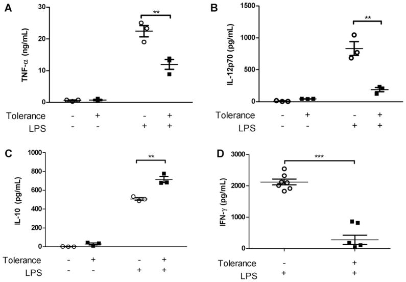
Results: Tolerized BM-DCs exhibited a significant drop in TNF-α and IL-12p70 production and increased IL-10 expression compared to untolerized cells. BM-DCs also showed the ability to develop heterotolerance, in which the LPS exposure alone was able to induce tolerance to Helicobacter pylori sonicate and TLR2 agonist Pam3Cys. Furthermore, the expression of IRAK-M was increased after restimulation of tolerized BM-DCs as determined qPCR and Western blot. IRAK-M exhibited a suppressive effect on surface expression of major histocompatibilty complex class II (MHC II) and CD80 in LPS-tolerized BM-DCs. IL-10 expression in bacterial sonicate-tolerized IRAK-M-/- BM-DCs was altered as compared to wild type BM-DCs, with tolerance-induced expression of IL-10 mitigated in tolerized IRAK-M-/- BM-DCs.

Conclusion: Along with endotoxin, bacterial sonicate is able to induce refractory tolerance in BM-DCs, and IRAK-M plays a role in modulating cell surface expression of MHC class II and CD80 and release of IL-10 during this tolerance.


Conflict of interest statement

Conflicts of interest

There is no conflict of interest declared for any author.

Figures

Fig. 1
Fig. 1
Low-dose LPS induces tolerance of BM-DCs to high-dose LPS re-stimulation. BM-DCs exposed to low dose (10 ng/ml) LPS (tolerized group) or PBS (non-tolerized group), washed in PBS, rested overnight, was re-stimulated by higher dose LPS (100 ng/ml). (A and B) BM-DC in the tolerance group exhibit a significant ablation in secreted TNF-α and IL-12p70 compared to BM-DCs in the No-tolerance group. (C) BM-DCs in the tolerized group show a significant increase in secreted IL-10 upon second stimulation by higher dose LPS compared to DCs not exposed to low dose LPS. (D) High-dose LPS re-stimulated BM-DCs were co-cultured with syngeneic splenocytes for 72 h in a mixed leukocyte reaction and the level of IFN-γ was measured by ELISA. Tolerized BM-DCs show an inability to stimulate a type I helper T cell response (Th1) compared to BM-DCs in the non-tolerized group. All cytokine measurements determined by ELISA. 1st (low)/2nd (high) dose LPS = 10/100 ng/ml or PBS. Data represent mean ± SEM, n = 3 in duplicates (n = 7 for mixed leukocyte reaction). *p < 0.05, **p < 0.01, ***p < 0.001 compared to untolerized cells.
Fig. 2
Fig. 2
TNF-α, IL-12p70 and IL-10 expression in supernatant of DCs tolerized and stimulated with H. pylori SS1 sonicate or E. coli sonicate. (A and B) When tolerized with E. coli or H. pylori sonicate, BM-DCs exhibit a significant ablation in secreted TNF-α and IL-12p70 upon second stimulation by a higher dose of the same sonicate as compared to non-tolerized cells. Between stimulations cells are washed with PBS and rested overnight. Non-tolerized DCs display a clear TNF-α and IL-12p70 response to sonicate stimulation. (C) Sonicate tolerization leads to a significant increase in secreted IL-10 upon second stimulation by higher dose H. pylori sonicate or E. coli sonicate as compared to non-tolerized BM-DCs. All cytokine measurements determined by ELISA. 1st (low)/2nd (high) dose sonicate = 2/10 μg/ml or PBS. Data represent mean ± SEM, n = 3 in duplicates. *p < 0.05, **p < 0.01, ***p < 0.001 compared to untolerized cells.
Fig. 3
Fig. 3
Heterotolerance in DCs exposed to low-dose LPS and subsequently exposed to H. pylori SS1 sonicate or TLR2 ligand. BM-DCs exposed to low-dose LPS (10 ng/ml), washed with PBS, rested overnight, and then exposed to H. pylori SS1 (10 μg/ml) sonicate or TLR2 ligand (Pam3Cys, EMC microcollections, Tubingen, Germany, 100 ng/ml). BM-DCs from the tolerized group exhibit a similar cytokine expression as homotolerized cells (exposed to a stimulus at a low dose and re-stimulated with the same stimulus at a higher dose) or PBS. Heterotolerized cells show ablated TNF-β expression and/or IL-12 and increased IL-10 expression when subjected first to low dose LPS then H. pylori sonicate or Pam3Cys compared to no-tolerance group BM-DCs. All cytokine measurements determined by ELISA. Data represent mean ± SEM, n = 3 (for H. pylori group) or 4 (for Pam3Cys group) in duplicates. *p < 0.05, **p < 0.01, ***p < 0.001 compared to untolerized cells.
Fig. 4
Fig. 4
IRAK-M mRNA and protein expression in endotoxin-tolerized wild type BM-DCs (A) qPCR 3 h after 2nd stimulation. BM-DCs were exposed for 8 h to low-dose LPS (10 ng/ml), washed with PBS, rested overnight, and re-stimulated with high-dose LPS (100 ng/ml). Cells were lysed and mRNA purified and copied to produce cDNA for qPCR analysis. After exposure to low-dose LPS there is a significantly increased production of IRAK-M transcript upon second stimulation by a higher dose of LPS. Data normalized to housekeeping gene GAPDH. (B) Western blot 8 h after re-stimulation with high-dose LPS. BM-DCs exposed to low dose LPS, washed with PBS, rested overnight, and re-stimulated with high dose LPS show increased intracellular IRAK-M expression compared to cells not exposed to low dose LPS. 1st (low)/2nd (high) dose LPS = 10/100 ng/ml or PBS. Data represent mean ± SEM, n = 3 in duplicates. *p < 0.05, **p < 0.01.
Fig. 5
Fig. 5
IRAK-M inhibits surface expression of MHC class II and CD80 in LPS-tolerized BM-DCs. BM-DCs were tolerized with LPS and then re-stimulated with PBS or LPS. The expression of major histocompatibility complex class II (MHC II) and co-stimulatory molecule CD80 were measured by FACS. Parts (A) and (B) are single-stained cells (for either MHC II or CD80), with an arbitrary intensity threshold to determine positive or negative expression, while part (C) is a representative plot of double-stained cells (for both MHC II and CD80) gated for double-positive wild type BM-DCs. (A) On initial stimulation and without further stimulation after tolerance, the expression of MHC II is significantly higher in IRAK-M−/− LPS-tolerized BM-DCs as compared to wild type LPS-tolerized BM-DCs. After LPS re-stimulation, further increase in MHC II was observed in IRAK-M−/− LPS-tolerized BM-DCs but not in wild type LPS-tolerized BM-DCs. (B) The pattern of expression of CD80 in IRAK-M−/− and wild type LPS-tolerized BM-DCs was found to be similar to that of MHC II, however only during tolerance are significant increases found in IRAK-M−/− cells as compared to wild type. (C) Representative flow plot demonstrates increase of MHC II and CD80 double-positive cells in IRAK-M−/− BM-DCs compared to wild type with gating held constant. Largest population of MHC II and CD80 double-positive cells is seen in tolerized IRAK-M−/− BM-DCs that have been re-stimulated with LPS. 1st (low)/2nd (high) dose LPS = 10/100 ng/ml or PBS. Data represent mean ± SEM, n = 3 in duplicates. *p < 0.05, **p < 0.01, ***p < 0.001.
Fig. 6
Fig. 6
TNF-α, IL-12p70 and IL-10 expression in supernatant of wild type vs IRAK-M−/− DCs tolerized with LPS. (A and B) Tolerized BM-DCs exhibit a significant ablation in secreted TNF-α and IL-12p70 upon second stimulation by higher dose LPS in both IRAK-M−/− or wild type mice. (C) IL-10 expression in BM-DCs was increased in both tolerized IRAK-M−/− BM-DCs and wild type BM-DCs compared to untolerized cells of the same type. Fold change indicates the change in cytokine expression when cells are tolerized and compared to non-tolerized cells of the same type, either wild type or IRAK-M−/−. An asterisk (*) after the IRAK-M−/− fold change indicates the change as being significantly different as compared to wild type cells. All cytokine measurements determined by ELISA. 1st (low)/2nd (high) dose LPS = 10/100 ng/ml or PBS. Data represent mean ± SEM, n = 3 in duplicates. *p < 0.05, **p < 0.01, ***p < 0.001 compared to untolerized cells.
Fig. 7
Fig. 7
TNF-α, IL-12p70 and IL-10 in supernatant of wild type compared to IRAK-M−/− DCs tolerized with H. pylori sonicate or E. coli sonicate. (A and B, D and E) BM-DCs tolerized with E. coli or H. pylori sonicate exhibit a significant decrease in secreted TNF-α and IL-12p70 upon second stimulation by higher dose E. coli or H. pylori sonicate compared to untolerized cells from both IRAK-M−/− or wild type mice. (C and F) IL-10 demonstrates mitigated increase during tolerance by the lack of IRAK-M. Tolerized wild type BM-DCs show an expected IL-10 increase upon re-stimulation, however tolerized IRAK-M−/− BM-DCs failed to significantly increase IL-10 expression as compared to untolerized IRAK-M−/− BM-DCs. Fold change indicates the change in cytokine expression when cells are tolerized and compared to non-tolerized cells of the same type, either wild type or IRAK-M−/−. An asterisk (*) after the IRAK-M−/− fold change indicates the change as being significantly different as compared to wild type cells. All cytokine measurements determined by ELISA. 1st (low)/2nd (high) dose sonicate = 2/10 μg/ml or PBS. Data represent mean ± SEM, n = 3 in duplicates. *p < 0.05, **p < 0.01, ***p < 0.001 compared to untolerized cells of the same genotype.

References

    1. Cherayil BJ. How not to get bugged by bugs: mechanisms of cellular tolerance to microorganisms. Curr Opin Gastroenterol. 2003;19:572–7. - PubMed
    1. Palucka K, Banchereau J. Dendritic cells: a link between innate and adaptive immunity. J Clin Immunol. 1999;19:12–25. - PubMed
    1. Liu ZJ, Yan LN, Li XH, Xu FL, Chen XF, You HB, et al. Up-regulation of IRAK-M is essential for endotoxin tolerance induced by a low dose of lipopolysaccharide in Kupffer cells. J Surg Res. 2008;150:34–9. - PubMed
    1. del Fresno C, Garcia-Rio F, Gomez-Pina V, Soares-Schanoski A, Fernandez-Ruiz I, Jurado T, et al. Potent phagocytic activity with impaired antigen presentation identifying lipopolysaccharide-tolerant human monocytes: demonstration in isolated monocytes from cystic fibrosis patients. J Immunol. 2009;182:6494–507. - PubMed
    1. Deng JC, Cheng G, Newstead MW, Zeng X, Kobayashi K, Flavell RA, et al. Sepsis-induced suppression of lung innate immunity is mediated by IRAK-M. J Clin Invest. 2006;116:2532–42. - PMC - PubMed

Publication types

MeSH terms