Comparative Study
. 2012 Apr 24;106(9):1512-9.
doi: 10.1038/bjc.2012.126.

Dynamic equilibrium between cancer stem cells and non-stem cancer cells in human SW620 and MCF-7 cancer cell populations

Affiliations
Comparative Study

Dynamic equilibrium between cancer stem cells and non-stem cancer cells in human SW620 and MCF-7 cancer cell populations

G Yang et al. Br J Cancer. .

Abstract

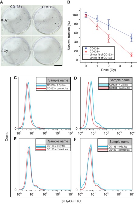
Background: Cancer stem cells (CSCs) paradigm suggests that CSCs might have important clinical implications in cancer therapy. Previously, we reported that accumulation efficiency of CSCs is different post low- and high-LET irradiation in 48 h.

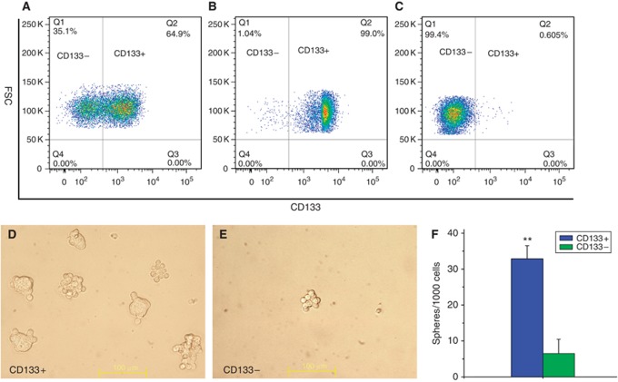
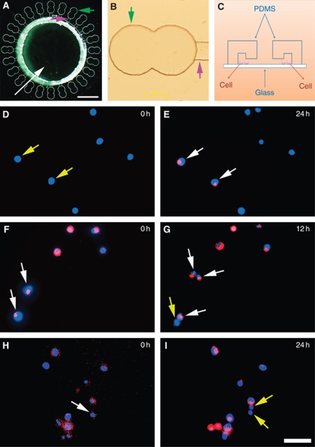
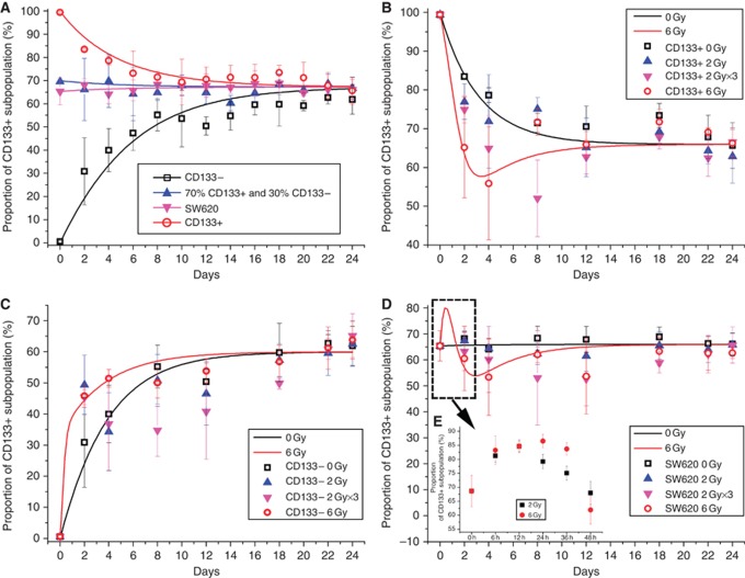
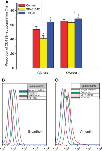
Methods: Cancer stem cells and non-stem cancer cells (NSCCs) were sorted and functionally identified through a variety of assays such as antigen profiles and sphere formation. Inter-conversion between CSCs and NSCCs were in situ visualised. Cancer stem cells proportions were assayed over multiple generations under normal and irradiation surroundings. Supplement and inhibition of TGF-β1, as well as immunofluorescence assay of E-cadherin and Vimentin, were performed.

Results: Surface antigen markers of CSCs and NSCCs exist in an intrinsic homoeostasis state with spontaneous and in situ visualisable inter-conversions, irrespective of prior radiations. Supplement with TGF-β1 accelerates the equilibrium, whereas inhibition of TGF-β signalling disturbs the equilibrium and significantly decreases CSC proportion. Epithelial mesenchymal transition (EMT) might be activated during the process.

Conclusion: Our results indicate that the intrinsic inter-conversion and dynamic equilibrium between CSCs and NSCCs exist under normal and irradiation surroundings, and TGF-β might have important roles in the equilibrium through activating EMT.


Figures

Figure 1
Figure 1
Enrichment of CD133+ CSCs and CD133 NSCCs through FACS. (AC) typical image of FACS sorting and efficiency. (A) Sorting of CD133+ and CD133 cell subpopulation. (B) Typical enrichment efficiency of CD133+ subpopulation from A. (C) Typical enrichment efficiency of CD133 subpopulation from A. (DF) Sphere formation of CD133+ and CD133 cells. (D) Typical image of sphere formations of CD133+ cells. (E) Typical image of sphere formations of CD133 cells, bar=100 μm. (F) Statistics in (D and E) at day 4 post inoculation (mean±s.d., n=3; **P=0.00109).
Figure 2
Figure 2
Radioresistance of CSCs. (A and B) Colony formation of CD133+ CSCs and NSCCs with or without irradiation. (A) Representative colony formation images of CD133+ CSC and CD133 NSCC controls (upper panels) and cultures irradiated with a 2-Gy dose of γ-rays (lower panels), bar=10 mm. (B) Survival fractions of CD133+ CSCs and CD133 NSCCs as a function of radiation dose. Each data point represents mean±s.d., n⩾3. (CF) γ-H2AX assay of CD133+ CSCs and CD133 NSCCs with or without irradiation. (C) Representative immunofluorescence image of γ-H2AX analysed by flow cytometry of CD133 NSCCs with or without a 2-Gy dose of γ-rays. (D) Similar analyses of CD133 NSCCs with or without a 4-Gy dose of γ-rays. (E) Analyses of CD133+ CSCs irradiated with 2 Gy and of controls. (F) Analyses of CD133+ CSCs irradiated at 4 Gy and of controls.
Figure 3
Figure 3
In situ immunofluorescence of CD133 expressions. (AC) Constructed microfluidic chip for in situ immunofluorescence of CD133 expressions. (A) The planform of a typical chip, white arrow indicates the reservoir, green arrow indicates the cell culture room of the chip, and purple arrow indicates the micro-channel of the chip, bar=1 mm. (B) The cell culture room (indicated as green arrow) and the channel (indicated as purple arrow) of a typical microfluidic chip, bar=200 μm. (C) The schematic diagram of a microfluidic chip. (D and E) Typical in situ transition of CD133+ CSCs from CD133 NSCCs (indicated as white arrows in E). (F and G) Typical CSC self-renewal (indicated as two white arrows in G) and asymmetric division (indicated as one white and one yellow arrow in G). (H and I) Typical differentiation of CSCs into NSCCs (indicated as two white arrow in I), bar=50 μm for DI. Yellow arrows indicate NSCCs and white arrows indicate CSCs. Images were merged with nuclei and CD133 expression patterns through Image-Pro Plus software (Media Cybernetics, Bethesda, MD, USA).
Figure 4
Figure 4
Intrinsic homoeostasis between CD133+ CSCs and CD133 NSCCs. (A) Dynamic proportion of CSC subpopulation of purified CSCs, NSCCs, mixture of the two cell types, and unsorted SW620 controls over a period of 24 days. (B) Dynamic proportion of CSC subpopulation in purified CD133+ cells with or without irradiation. (C) Dynamic proportion of CSC subpopulation in purified CD133 NSCCs with or without irradiation. (D) Dynamic proportion of CSC subpopulation in unsorted SW620 cells with or without irradiation. (E) Details of data from the bracketed curve covering 0–48 h. Each data point represents mean±s.d., n⩾3.
Figure 5
Figure 5
TGF-β1 might have important roles in the inter-conversion and homoeostasis between NSCCs and CSCs via activating EMT. (A) Exogenous supplement of TGF-β1 accelerates transition of CSCs from NSCCs (mean±s.d., n=3; *P=0.02982), whereas SB431542 inhibits the transition (mean±s.d., n=3; *P=0.02395); TGF-β1- and SB431542-treated unsorted SW620 cells showed a significant difference in CSC proportion (mean±s.d., I=3; *P=0.03757). (B) Expressions of E-cadherin in purified NSCCs with or without TGF-β1 and SB431542. (C) Expressions of Vimentin in purified NSCCs with or without TGF-β1 and SB43154. Data was detected through FACS at day 8 post inoculation.

Similar articles

Cited by

References

    1. Achuthan S, Santhoshkumar TR, Prabhakar J, Nair SA, Pillai MR (2011) Drug-induced senescence generates chemoresistant stemlike cells with low reactive oxygen species. J Biol Chem 286(43): 37813–37829 - PMC - PubMed
    1. Adams JM, Kelly PN, Dakic A, Nutt SL, Strasser A (2007) Response to comment on ‘tumor growth need not be driven by rare cancer stem cells’. Science 318(5857): 1722 - PubMed
    1. Al-Hajj M, Wicha MS, Benito-Hernandez A, Morrison SJ, Clarke MF (2003) Prospective identification of tumorigenic breast cancer cells. Proc Natl Acad Sci USA 100(7): 3983–3988 - PMC - PubMed
    1. Alison MR, Lim SML, Nicholson LJ (2011) Cancer stem cells: problems for therapy? J Pathol 223(2): 148–162 - PubMed
    1. Andarawewa KL, Erickson AC, Chou WS, Costes SV, Gascard P, Mott JD, Bissell MJ, Barcellos-Hoff MH (2007) Ionizing radiation predisposes nonmalignant human mammary epithelial cells to undergo transforming growth factor beta induced epithelial to mesenchymal transition. Cancer Res 67(18): 8662–8670 - PubMed

Publication types

MeSH terms