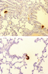
Dynamics of calcitonin gene-related peptide-like cells changes in the lungs of two-kidney, one-clip rats
- PMID: 22472888
- PMCID: PMC3352129
- DOI: 10.4081/ejh.2012.e10
Dynamics of calcitonin gene-related peptide-like cells changes in the lungs of two-kidney, one-clip rats
Abstract
Taking into consideration renal hypertension-induced homeostatic disorders and the key role of calcitonin gene-related peptide (CGRP) in many, systemic functions regulating systems, a question arises as to what an extent arterial hypertension affects the morphology and dynamics of pulmonary CGRP-immunopositive cell changes. The aim of the present study was to examine the distribution, morphology and dynamics of changes of CGRP-containing cells in the lungs of rats in the two-kidney, one-clip (2K1C) renovascular hypertension model. The studies were carried out on the lungs of rats after 3, 14, 28, 42, and 91 days long period from the renal artery clipping procedure. In order to identify neuroendocrine cells, immunohistochemical reaction was performed with the use of a specific antibody against CGRP. It was revealed that renovascular hypertension caused changes in the neuroendocrine, CGRP-containing cells in the lungs of rats. The changes, observed in the neuroendocrine cells, depended on time periods from experimentally induced hypertension. The highest intensity of changes in the neuroendocrine cells was observed in the lungs of rats after 14 days from the surgery.
Figures


Similar articles
-
Quantitative distribution and localization of calcitonin gene-related peptide-like cells in the stomach of two kidney, one clip rats.J Physiol Pharmacol. 2009 Jun;60(2):35-9. J Physiol Pharmacol. 2009. PMID: 19617643
-
Dynamics of cocaine- and amphetamine-regulated transcript containing cell changes in the adrenal glands of two kidney, one clip rats.Exp Biol Med (Maywood). 2014 Oct;239(10):1292-9. doi: 10.1177/1535370214538593. Epub 2014 Jun 17. Exp Biol Med (Maywood). 2014. PMID: 24939825
-
An increase in the synthesis and release of calcitonin gene-related peptide in two-kidney, one-clip hypertensive rats.Regul Pept. 2003 Jul 15;114(2-3):175-82. doi: 10.1016/s0167-0115(03)00124-1. Regul Pept. 2003. PMID: 12832107
-
Calcitonin gene-related peptide: role in airway homeostasis.Curr Opin Pharmacol. 2004 Jun;4(3):215-20. doi: 10.1016/j.coph.2004.01.006. Curr Opin Pharmacol. 2004. PMID: 15140411 Review.
-
Calcitonin gene-related peptide and hypertension.Peptides. 2005 Sep;26(9):1676-85. doi: 10.1016/j.peptides.2005.02.002. Epub 2005 Mar 2. Peptides. 2005. PMID: 16112410 Review.
Cited by
-
Influence of renovascular hypertension on the distribution of vasoactive intestinal peptide in the stomach and heart of rats.Exp Biol Med (Maywood). 2015 Nov;240(11):1402-7. doi: 10.1177/1535370215587533. Epub 2015 May 19. Exp Biol Med (Maywood). 2015. PMID: 25990439 Free PMC article.
-
Quantitative evaluation of CART-containing cells in urinary bladder of rats with renovascular hypertension.Eur J Histochem. 2015 Apr 13;59(2):2446. doi: 10.4081/ejh.2015.2446. Eur J Histochem. 2015. PMID: 26150151 Free PMC article.
References
-
- Przewlocki T, Kabłak-Ziembicka A, Tracz W, Kozanecki A, Kopec G, Rubis P, et al. Renal artery stenosis in patients with coronary artery disease. Kardiol Pol. 2008;66:856–62. - PubMed
-
- Imiela J. Renal artery stenosis renovascular hypertension ischaemic nephropathy. Kardiologia na co Dzien. 2009;4:21–7.
-
- Kokot F, Kokot J. Renovascular hypertension – a persistently difficult diagnostic problem. Post Hig Dośw. 1994;48:645–61. - PubMed
-
- Dzielińska Z, Januszewicz A, Demkow M, Makowiecka-Cieśla M, Prejbisz A, Naruszewicz M, et al. Cardiovascular risk factors in hipertensive patients with coronary artery disease and coexisting renal artery stenosis. J Hypertens. 2007;25:663–70. - PubMed
-
- Zellert T. Renal artery stenosis. Curr Treat Options Cardiovasc Med. 2007;9:90–9. - PubMed
MeSH terms
Substances
LinkOut - more resources
Full Text Sources
Research Materials

