

Histochemical localization of sialic acids and antimicrobial substances in eccrine glands of porcine snout skin
- PMID: 22472894
- PMCID: PMC3352135
- DOI: 10.4081/ejh.2012.e6
Histochemical localization of sialic acids and antimicrobial substances in eccrine glands of porcine snout skin
Abstract
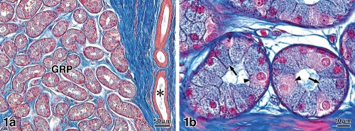
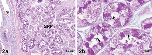
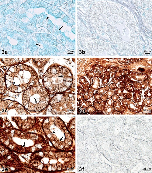
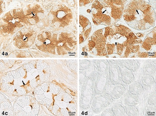
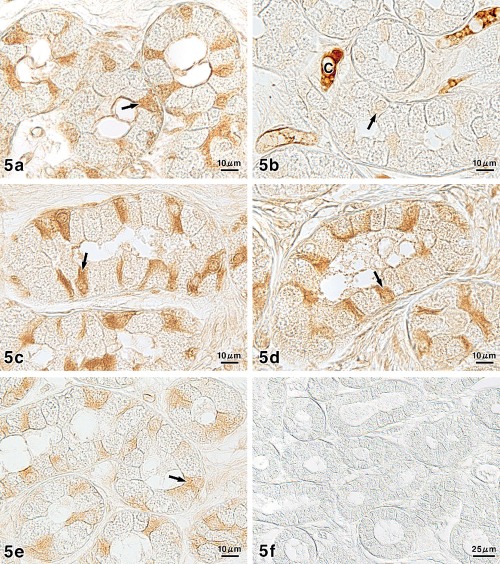
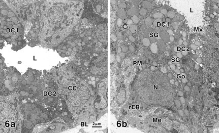
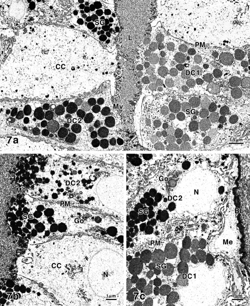
The distribution of sialic acids and antimicrobial products (lysozyme, IgA, lactoferrin, β-defensin 2) as well as Rab3D in the eccrine glands of porcine snout skin was studied by sialoglycoconjugate histochemistry and immunohistochemistry. The secretory epithelium consisted of two types of secretory cells: dark and clear cells. The dark cells exhibited considerable amounts of sialoglycoconjugates, which included O-acetylated sialic acids, whereas sialic acids in the sequence Siaα2-3Gal1-4GlcNAc were confined to some of the dark cells. All antimicrobial substances and Rab3D were demonstrated to be also mainly present in some of the dark cells. Additionally, in the cytological and cytochemical features, the different characteristics were observed among the dark cells. The results obtained are discussed with regard to the functional significance of the eccrine glands. The secretory products elaborated by this gland type may function as protective agents in order to preserve the skin integrity of the snout region, considering that sialic acids and antimicrobial substances are important in general defense mechanisms.
Figures
Similar articles
-
Histochemical distribution of sialic acids and antimicrobial substances in porcine carpal glands.Arch Dermatol Res. 2012 Oct;304(8):599-607. doi: 10.1007/s00403-012-1226-4. Epub 2012 Mar 20. Arch Dermatol Res. 2012. PMID: 22426985
-
Histochemical properties of sialic acids and antimicrobial substances in canine anal glands.Eur J Histochem. 2011;55(3):e29. doi: 10.4081/ejh.2011.e29. Epub 2011 Aug 27. Eur J Histochem. 2011. PMID: 22073376 Free PMC article.
-
Cytochemistry of sialoglycoconjugates, lysozyme, and β-defensin in eccrine glands of porcine snout skin as studied by electron microscopy.Microsc Res Tech. 2013 Jan;76(1):12-9. doi: 10.1002/jemt.22129. Epub 2012 Oct 3. Microsc Res Tech. 2013. PMID: 23032992
-
Defensins in saliva and the salivary glands.Med Electron Microsc. 2003 Dec;36(4):247-52. doi: 10.1007/s00795-003-0225-0. Med Electron Microsc. 2003. PMID: 16228657 Review.
-
Avian antimicrobial peptides: the defense role of beta-defensins.Biochem Biophys Res Commun. 2004 Oct 22;323(3):721-7. doi: 10.1016/j.bbrc.2004.08.162. Biochem Biophys Res Commun. 2004. PMID: 15381059 Review.
Cited by
-
On the future contents of a small journal of histochemistry.Eur J Histochem. 2012 Dec 10;56(4):e51. doi: 10.4081/ejh.2012.e51. Eur J Histochem. 2012. PMID: 23361247 Free PMC article.
-
Insulin granule morphology and crinosome formation in mice lacking the pancreatic β cell-specific phogrin (PTPRN2) gene.Histochem Cell Biol. 2024 Mar;161(3):223-238. doi: 10.1007/s00418-023-02256-8. Epub 2023 Dec 27. Histochem Cell Biol. 2024. PMID: 38150052
-
Exploration of the Sialic Acid World.Adv Carbohydr Chem Biochem. 2018;75:1-213. doi: 10.1016/bs.accb.2018.09.001. Epub 2018 Nov 28. Adv Carbohydr Chem Biochem. 2018. PMID: 30509400 Free PMC article. Review.
-
Ultrastructure and immunohistochemical characterization of proteins concerned with the secretory machinery in goat ceruminous glands.Eur J Histochem. 2017 Aug 8;61(3):2828. doi: 10.4081/ejh.2017.2828. Eur J Histochem. 2017. PMID: 29046053 Free PMC article.
References
-
- Ellis RA. Eccrine sweat glands: electron microscopy, cytochemistry and anatomy. In: Jadassohn J, editor. Handbuch der Haut- und Geschlechtskrankheiten, Ergänzungswerk I/1. Springer; Berlin, Germany: 1968. pp. 224–66.
-
- Calhoun ML, Stinson AW. Integument. In: Dellman HD, Brown EM, editors. Textbook of veterinary histology. 2nd edn. Lea and Febiger; Philadelphia, USA: 1981. pp. 378–411.
-
- Tsukise A, Fujimori O, Yamada K. Histochemistry of glycoconjugates in the goat nasolabial skin with special reference to eccrine glands. Acta Anat. 1988;132:150–8. - PubMed
-
- Tsukise A, Meyer W, Fujimori O, Yamada K. The cytochemistry of glycoconjugates in the planum nasolabial glands of the goat as studied by electron microscopic methods. Histochem J. 1988;20:617–23. - PubMed
-
- Meyer W, Bartels T. Histochemical study on the eccrine glands in the foot pad of the cat. Basic Appl Histochem. 1989;33:219–38. - PubMed
MeSH terms
Substances
LinkOut - more resources
Full Text Sources
Miscellaneous
