

NADPH-d activity in rat thymus after the application of retinoid acid
- PMID: 22472895
- PMCID: PMC3352136
- DOI: 10.4081/ejh.2012.e7
NADPH-d activity in rat thymus after the application of retinoid acid
Abstract
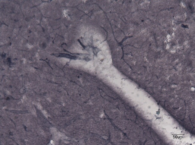
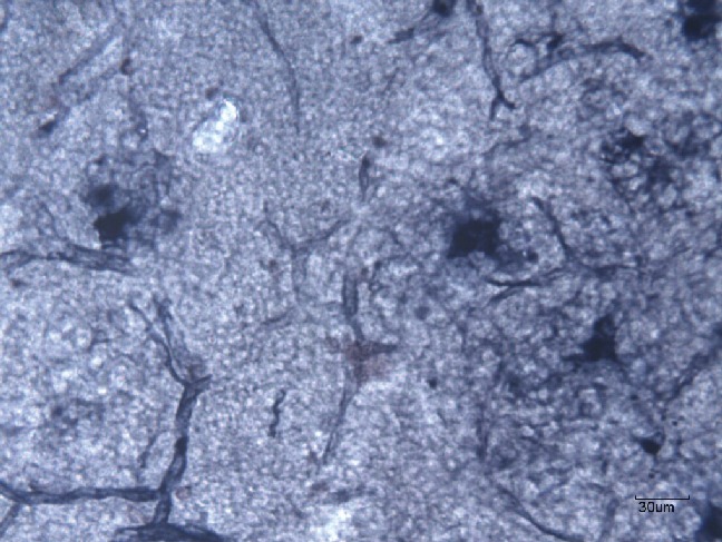
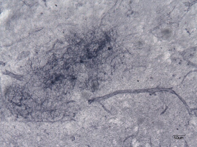
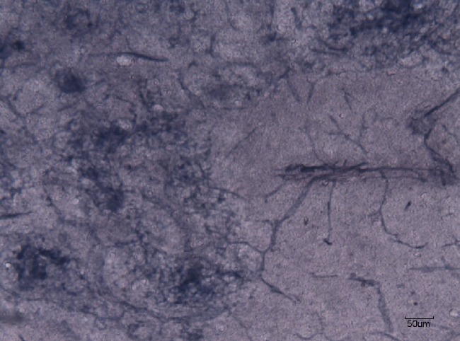
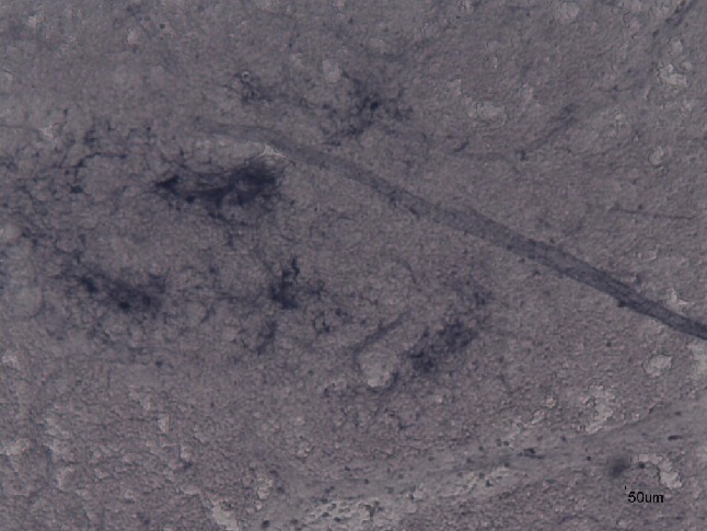
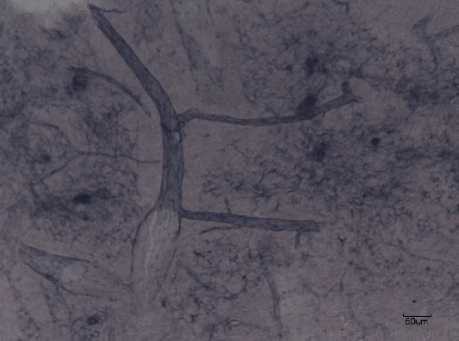
The aim of this work was to determine the localization of nicotinamide-adenine dinucleotide phosphate-diaphorase (NADPH-d) activity as the marker for synthesis of nitric oxide synthase (NOS) in the rat thymus after the application of retinoid acid (RA) on 1st, 7th, 14th and 21st days of gestation. The given results can build the basis for understanding of the role of NOS in rat thymus. NADPH-d positive cells were represented with dark-blue color and were localized on corticomedullar junction of the thymus. These cells were of different intensity of coloring and were shaped in oval, circle or irregular forms. NADPH-d positive nerve fibers were observed in perivascular topography. They were marked more strongly in the case of control group. The result of application of RA to gravid rats was that the birth weights of newborn rats and their thymuses were smaller, but without statistically significance.
Figures
Similar articles
-
Localisation of NADPH-diaphorase-positive structures in the thymus of the rat, mouse and rabbit.Folia Morphol (Warsz). 2003;62(3):167-70. Folia Morphol (Warsz). 2003. PMID: 14507040
-
[Localization of NADPH-d positive structures in the thymus of rabbits and rats].Bratisl Lek Listy. 1997 Feb;98(2):102-6. Bratisl Lek Listy. 1997. PMID: 9264807 Slovak.
-
[Localization of NADPH-d positive cells in the thymus of pheasants and mice].Bratisl Lek Listy. 1998 Feb;99(2):104-7. Bratisl Lek Listy. 1998. PMID: 9588087 Slovak.
-
Comparative study of the topographic localization of NADPH diaphorase positive cells in the rabbit and pheasant thymuses.Ukr Biokhim Zh (1999). 2002 Jan-Feb;74(1):111-6. Ukr Biokhim Zh (1999). 2002. PMID: 12199090
-
Ultrastructure of NADPH diaphorase-positive nerve fibers and their terminals in the rat cerebral arterial system.J Chem Neuroanat. 2001 Jun;21(4):267-75. doi: 10.1016/s0891-0618(01)00090-4. J Chem Neuroanat. 2001. PMID: 11429268
Cited by
-
Histochemistry as an irreplaceable approach for investigating functional cytology and histology.Eur J Histochem. 2013 Dec 19;57(4):e41. doi: 10.4081/ejh.2013.e41. Eur J Histochem. 2013. PMID: 24441194 Free PMC article.
-
Betulinic acid‑induced expression of nicotinamide adenine dinucleotide phosphate‑diaphorase in the immune organs of mice: A possible role of nitric oxide in immunomodulation.Mol Med Rep. 2018 Feb;17(2):3035-3041. doi: 10.3892/mmr.2017.8262. Epub 2017 Dec 12. Mol Med Rep. 2018. PMID: 29257292 Free PMC article.
-
Impact of Histochemistry on biomedical research: looking through the articles published in a long-established histochemical journal.Eur J Histochem. 2014 Dec 30;58(4):2474. doi: 10.4081/ejh.2014.2474. Eur J Histochem. 2014. PMID: 25578981 Free PMC article.
References
-
- DeLuca LM, Darwiche N, Jones CS, Scita G. Retinoids in differentiation and neoplasia. Sci Am Sci Med. 1995;2:28–37.
-
- Zile MH. Vitamin A and embryonal development. J Nutr. 1998;128:4555–85. - PubMed
-
- Hale F. Pigs born without eyeballs. J Heredit. 1933;24:105–06.
-
- Cohlan SQ. Excessive intake of vitamin A as a cause of congenital anomalies in the rat. Science. 1953;117:535–36. - PubMed
-
- Velíšek J, Hajšlová V. Chemie potravin [Article in Czech] Tabor. 2009
Publication types
MeSH terms
Substances
LinkOut - more resources
Full Text Sources
