. 2012 Jun;20(6):1187-95.
doi: 10.1038/mt.2012.59. Epub 2012 Apr 3.

Lentiviral vector induced insertional haploinsufficiency of Ebf1 causes murine leukemia

Affiliations

Lentiviral vector induced insertional haploinsufficiency of Ebf1 causes murine leukemia

Dirk Heckl et al. Mol Ther. 2012 Jun.

Abstract

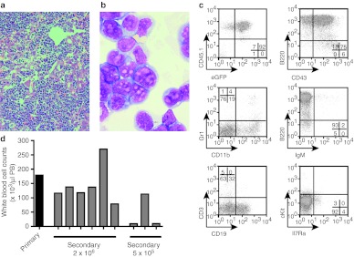
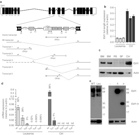
Integrating vectors developed on the basis of various retroviruses have demonstrated therapeutic potential following genetic modification of long-lived hematopoietic stem and progenitor cells. Lentiviral vectors (LV) are assumed to circumvent genotoxic events previously observed with γ-retroviral vectors, due to their integration bias to transcription units in comparison to the γ-retroviral preference for promoter regions and CpG islands. However, recently several studies have revealed the potential for gene activation by LV insertions. Here, we report a murine acute B-lymphoblastic leukemia (B-ALL) triggered by insertional gene inactivation. LV integration occurred into the 8th intron of Ebf1, a major regulator of B-lymphopoiesis. Various aberrant splice variants could be detected that involved splice donor and acceptor sites of the lentiviral construct, inducing downregulation of Ebf1 full-length message. The transcriptome signature was compatible with loss of this major determinant of B-cell differentiation, with partial acquisition of myeloid markers, including Csf1r (macrophage colony-stimulating factor (M-CSF) receptor). This was accompanied by receptor phosphorylation and STAT5 activation, both most likely contributing to leukemic progression. Our results highlight the risk of intragenic vector integration to initiate leukemia by inducing haploinsufficiency of a tumor suppressor gene. We propose to address this risk in future vector design.


Figures

Figure 1
Figure 1
Acute B-lymphoblastic leukemia. (a) Representative histology showing strong infiltration of leukemic cells in the liver. (H&E staining, ×200). (b) Leukemic blasts from the bone marrow of the primary leukemia present with lymphoid and myeloid characteristics. Most blasts cells are larger and have typical myeloid (monocytic) cellular outlines and cytoplasm. The nucleoli are larger and irregular, which are usually seen in lymphoblasts rather than in myeloblasts. (May-Grünwald/Giemsa, ×1,000). (c) Flow cytometry of primary leukemic BM cells indentified the leukemia as B-lymphoblastic with expression of B220 and CD43 while lacking IgM expression. CD19 was expressed on ~32% of the cells while cKit and IL7Ra expression was almost absent. Approximately 19% of the cells expressed the myelo-monocytic marker CD11b. CD45.1 and GFP expression marked cells as donor derived and vector transduced. (d) BM cells of the primary mouse that developed B-lymphoblastic leukemia were transplanted into lethally irradiated secondary recipient mice at two different doses: 2 × 10⁶ and 5 × 10⁵ BM cells. All mice that received the high cell dose succumbed to leukemia 12–17 days after transplantation with high peripheral white blood cell counts, whereas only one of the recipients that were transplanted with the low dose of cells developed leukocytosis. BM, bone marrow; eGFP, enhanced green fluorescent protein; H&E, hematoxylin and eosin; PB, peripheral blood.
Figure 2
Figure 2
B-ALL consist of a clone with two insertion sites. (a) Southern blot analysis of BM samples from different secondary recipients (lane 1–6) or control mice (7–9) digested with BsrGI (1, 2, 7), EcoNI (3, 4, 8) or NcoI (5, 6, 9) and probed for the vector-specific post-transcriptional regulatory element (PRE). (b) Ligation-mediated PCR (LM-PCR) on genomic DNA from the BM of a secondary recipient verified two vector integrations which were identified by sequencing to be in the Nhs gene and in the Ebf1 gene. BM, bone marrow.
Figure 3
Figure 3
Analysis of the genomic stability. (a) No chromosomal aberrations were detected by spectral karyotyping (SKY). Leukemic cell were grown in culture (IL7, Flt3L) for 2 days to induce proliferation and metaphases prepared. (b) Comparative genome hybridization (array-CGH) analysis of leukemic cells. Genomic profiles by means of high resolution array-CGH (180 k): Cye3/Cy5 log2 ratios of fluorescence intensities of probes against their chromosomal localization along chromosomes 1–19 is shown, X: stacked plots of genomic DNA samples of the secondary mice one (brown), two (green), three (black), and the primary mouse (blue); pooled DNA from 10 female C57BL/6 spleen specimen served as reference leading to monosomal X in the male test samples. (i) Microdeletion within chromosomal region 6 in all four probes ranging from 67.847–70.677 Mb containing no genes; (ii) microdeletion within chromosomal region 12 in all four probes ranging from 114.676–115.154 Mb containing no genes.
Figure 4
Figure 4
Post-transcriptional deregulation of Ebf1 following lentiviral vector insertion. (a) Schematic overview of the Ebf1 gene locus with the in-sense integrated SIN-LV in intron 8. The SIN-LV harbors the exon (E) and intron (I) containing GPIba promoter, the eGFP reporter gene, the PRE, and splice donors (SD), and splice acceptors (SA) sites as indicated. Splice events can lead to alternative transcripts as indicated in the figure (transcripts 1–5) in addition to the wt and vector transcripts. A stop codon can occur due to frame shifts in transcript 2. (b) Ebf1 mRNA expression levels relative to actin in the BM from three independent leukemic mice (leukemia) and three independently FACS-sorted CD19+CD43+ B-cell progenitor samples from wild-type mice BM (Ctrl). (c) Immunoblot analysis of Ebf1 expression in leukemic samples from bone marrow (BM), spleen (SP), and peripheral blood (PB) of leukemic mice in comparison to wild-type CD19+CD43+ B-cell progenitors (Ctrl). Actin was used as loading control. (d) Quantitative RT-PCR detecting the different splice products of the Ebf1 locus with the inserted LV in comparison to the full-length transcript. Readthrough transcripts into intron 8 (transcript 1) and fusion transcripts from exon 8 to the LV eGFP gene that result in a frame shift and early stop (transcript 2) are well-detectable. Splice products from the LV to the exon 9 (transcripts 3–5) could be amplified and sequences were verified (Supplementary Figure S6) but the overall amount of transcripts was extremely low (percentage of transcript in correlation to the full-length transcript is given). Results from three independent leukemic BM samples (leukemia) in comparison to pooled (n = 3) wild-type BM (Ctrl) (nd = not detected). (e) Immunoblot analysis of wild-type splenocytes (WT) and leukemic cells (Leu) does not show detectable amounts of truncated protein corresponding to the expected protein. Immunoblot analysis of 293T cells transfected with lentiviral constructs expressing dTomato alone (−), full-length Ebf1 (fl) or the truncated Ebf1 (tr) shows that the employed antibody was able to detect the truncated Ebf1 protein. eGFP, enhanced green fluorescent protein; FACS, fluorescence-activated cell sorting; LTR, long terminal repeat; mRNA, messenger RNA; pA, poly-A; PRE, post-transcriptional regulatory element; RT-PCR, reverse transcription-PCR; SIN-LV, self-inactivating lentiviral vector.
Figure 5
Figure 5
Expression of a truncated Ebf1 protein does not interfere with in vitro B-cell differentiation. Lineage negative BM cells were transduced with a SIN-lentiviral vector expressing the truncated variants of Ebf1, Ebf1-tr1 (RRL.PPT.SFFV.Ebf1-tr1.IRES.dTomato.pre) or Ebf1-tr2 (RRL.PPT.SFFV.Ebf1-tr2.IRES.dTomato.pre) which correspond to the truncations induced by readthrough or splicing to the GPIbaP splice acceptor, respectively. For detection by flow cytometry a dTomato was coexpressed from an IRES. As control, cells were transduced with a vector expressing dTomato alone (RRL.PPT.SFFV.IRES.dTomato.pre). (i) Transgene expression as indicated by dTomato expression in a representative culture on day 7 of coculture on OP9 stromal cells, (ii) expression of B-cell markers B220 and CD19 after 7 days, and (iii) after 18 days was assessed by flow cytometry. Percentages of the distinct populations are indicated in corresponding quadrants. No differences in B-cell differentiation were seen in cells expressing the truncated EBF1 protein in comparison to control vector-transduced cells.
Figure 6
Figure 6
Downregulation of Ebf1 target genes coincides with reactivation of myeloid genes and Csf1r upregulation. (a) cDNA-microarray analysis performed on three independent leukemic BM samples in comparison to three independent samples of CD19+CD43+ wild-type B-cell progenitors (wt B-cells). Gene set enrichment analysis (GSEA) shows downregulation of Ebf1 target genes in leukemic cells as compared to wild-type B-cell progenitors (NES = 1.55; P = 0.012, FDR <0.02). (b) GSEA analysis of myeloid lineage-specific genes shows strong enrichment of myeloid genes in leukemic cells (NES = −2.49; P < 0.001, FDR <0.001). (c) Phospho-tyrosine immunoblot analysis detected activation of STAT5 (Tyr694) in leukemic cells from bone marrow (BM), spleen (SP), and peripheral blood (PB) in comparison to wild-type CD19+CD43+ B-cell progenitors (Ctrl). (d) Strong phosphorylation of Csf1r [1] (aka. M-Csfr or c-Fms) and some minor activation of Pdgfr-β [2] was found by phospho-receptor-tyrosine-kinase protein arrays. Both genes had been found upregulated in leukemic cells in the cDNA-microarray analysis. Ctrl spots [3]. (e) Immunoblot analysis showing CSFR1, pSTAT5, STAT5, pERK 1/2, ERK 1/2, pAKT, and AKT expression/activation in leukemic cells grown on OP9 stromal cells with 20 ng/ml SCF, 20 ng/ml FLT3-L, and 10 ng/ml IL7 [1], starved for 3 hours [2], and stimulated with 40 ng/ml M-CSF for 10 minutes after 3 hours starvation [3]. Loss of STAT5 phosphorylation upon starvation and regain after stimulation with M-CSF shows ligand-dependency of upregulated CSF1R. FDR, false discovery rate; M-CSF, macrophage colony-stimulating factor; NES, normalized enrichment score.

Similar articles

Cited by

References

    1. Qasim W, Gaspar HB., and, Thrasher AJ. Progress and prospects: gene therapy for inherited immunodeficiencies. Gene Ther. 2009;16:1285–1291. - PubMed
    1. Stein S, Ott MG, Schultze-Strasser S, Jauch A, Burwinkel B, Kinner A.et al. (2010Genomic instability and myelodysplasia with monosomy 7 consequent to EVI1 activation after gene therapy for chronic granulomatous disease Nat Med 16198–204. - PubMed
    1. Howe SJ, Mansour MR, Schwarzwaelder K, Bartholomae C, Hubank M, Kempski H.et al. (2008Insertional mutagenesis combined with acquired somatic mutations causes leukemogenesis following gene therapy of SCID-X1 patients J Clin Invest 1183143–3150. - PMC - PubMed
    1. Hacein-Bey-Abina S, Garrigue A, Wang GP, Soulier J, Lim A, Morillon E.et al. (2008Insertional oncogenesis in 4 patients after retrovirus-mediated gene therapy of SCID-X1 J Clin Invest 1183132–3142. - PMC - PubMed
    1. Mitchell RS, Beitzel BF, Schroder AR, Shinn P, Chen H, Berry CC.et al. (2004Retroviral DNA integration: ASLV, HIV, and MLV show distinct target site preferences PLoS Biol 2E234. - PMC - PubMed

Publication types

MeSH terms