

Van Maldergem syndrome: further characterisation and evidence for neuronal migration abnormalities and autosomal recessive inheritance
- PMID: 22473091
- PMCID: PMC3449074
- DOI: 10.1038/ejhg.2012.57
Van Maldergem syndrome: further characterisation and evidence for neuronal migration abnormalities and autosomal recessive inheritance
Abstract
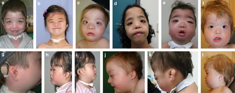
We present six patients from five unrelated families with a condition originally described by Van Maldergem et al and provide follow-up studies of the original patient. The phenotype comprises a distinctive facial appearance that includes blepharophimosis, maxillary hypoplasia, telecanthus, microtia and atresia of the external auditory meatus, intellectual disability, digital contractures and skeletal anomalies together with subependymal and subcortical neuronal heterotopia. Affected patients typically have neonatal hypotonia, chronic feeding difficulties and respiratory problems. In our cohort, we have observed one instance of sibling recurrence and parental consanguinity in three of the families, indicating that autosomal recessive inheritance is likely.
Figures


Similar articles
-
A newborn diagnosed with van Maldergem syndrome.Clin Dysmorphol. 2018 Apr;27(2):63-65. doi: 10.1097/MCD.0000000000000211. Clin Dysmorphol. 2018. PMID: 29505454 No abstract available.
-
A further patient with van Maldergem syndrome.Eur J Med Genet. 2012 Jun;55(6-7):423-8. doi: 10.1016/j.ejmg.2012.02.012. Epub 2012 Mar 13. Eur J Med Genet. 2012. PMID: 22469822
-
Van Maldergem syndrome and Hennekam syndrome: Further delineation of allelic phenotypes.Am J Med Genet A. 2018 May;176(5):1166-1174. doi: 10.1002/ajmg.a.38652. Am J Med Genet A. 2018. PMID: 29681106
-
Acro-cardio-facial syndrome.Orphanet J Rare Dis. 2010 Sep 29;5:25. doi: 10.1186/1750-1172-5-25. Orphanet J Rare Dis. 2010. PMID: 20920258 Free PMC article. Review.
-
A novel syndrome with psychiatric features and review of malformation syndromes with psychiatric disorders.Am J Med Genet A. 2009 Feb 15;149A(4):713-21. doi: 10.1002/ajmg.a.32709. Am J Med Genet A. 2009. PMID: 19253384 Review.
Cited by
-
Fat4/Dchs1 signaling between stromal and cap mesenchyme cells influences nephrogenesis and ureteric bud branching.Development. 2015 Aug 1;142(15):2574-85. doi: 10.1242/dev.122630. Epub 2015 Jun 26. Development. 2015. PMID: 26116666 Free PMC article.
-
Zebrafish endochondral growth zones as they relate to human bone size, shape and disease.Front Endocrinol (Lausanne). 2022 Dec 6;13:1060187. doi: 10.3389/fendo.2022.1060187. eCollection 2022. Front Endocrinol (Lausanne). 2022. PMID: 36561564 Free PMC article. Review.
-
Coexistence of genetic conditions: exploring a possible relationship.Sudan J Paediatr. 2019;19(1):60-66. doi: 10.24911/SJP.106-1554459680. Sudan J Paediatr. 2019. PMID: 31384091 Free PMC article.
-
Definitions and classification of malformations of cortical development: practical guidelines.Brain. 2020 Oct 1;143(10):2874-2894. doi: 10.1093/brain/awaa174. Brain. 2020. PMID: 32779696 Free PMC article. Review.
-
Analysis of recent shared ancestry in a familial cohort identifies coding and noncoding autism spectrum disorder variants.NPJ Genom Med. 2022 Feb 21;7(1):13. doi: 10.1038/s41525-022-00284-2. NPJ Genom Med. 2022. PMID: 35190550 Free PMC article.
References
-
- Van Maldergem L, Wetzburger C, Verloes A, Fourneau C, Gillerot Y. Mental retardation with blepharo-naso-facial abnormalities and hand malformations: a new syndrome. Clin Genet. 1992;41:22–24. - PubMed
-
- Zampino G, Colosimo C, Balducci F, et al. Cerebro-facio-articular syndrome of Van Maldergem: confirmation of a new MR/MCA syndrome. Clin Genet. 1994;45:140–144. - PubMed
-
- des Portes V, Pinard JM, Billuart P, et al. A novel CNS gene required for neuronal migration and involved in X-linked subcortical laminar heterotopia and lissencephaly syndrome. Cell. 1998;92:51–61. - PubMed
-
- Parrini E, Ramazzotti A, Dobyns WB, et al. Periventricular heterotopia: phenotypic heterogeneity and correlation with Filamin A mutations. Brain. 2006;129:1892–1906. - PubMed
Publication types
MeSH terms
Supplementary concepts
LinkOut - more resources
Full Text Sources
Medical
Molecular Biology Databases
