Review
. 2012 Sep;8(9):529-43.
doi: 10.1038/nrendo.2012.36. Epub 2012 Apr 3.

Arterial calcification and bone physiology: role of the bone-vascular axis

Affiliations
Review

Arterial calcification and bone physiology: role of the bone-vascular axis

Bithika Thompson et al. Nat Rev Endocrinol. 2012 Sep.

Abstract

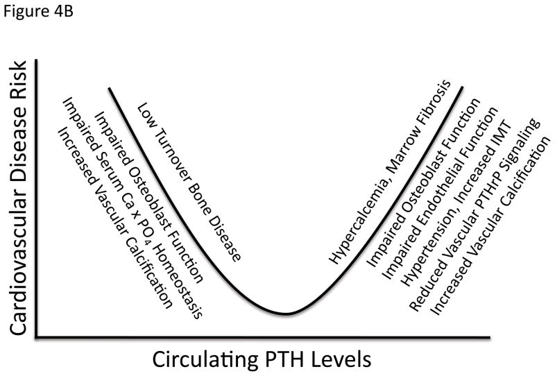
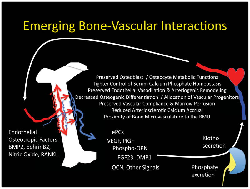
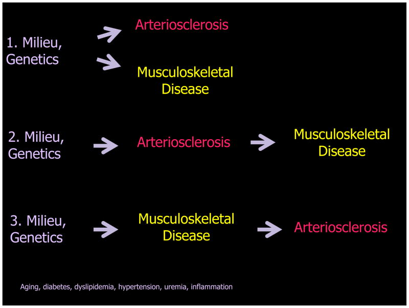
Bone never forms without vascular interactions. This simple statement of fact does not adequately reflect the physiological and pharmacological implications of the relationship. The vasculature is the conduit for nutrient exchange between bone and the rest of the body. The vasculature provides the sustentacular niche for development of osteoblast progenitors and is the conduit for egress of bone marrow cell products arising, in turn, from the osteoblast-dependent haematopoietic niche. Importantly, the second most calcified structure in humans after the skeleton is the vasculature. Once considered a passive process of dead and dying cells, vascular calcification has emerged as an actively regulated form of tissue biomineralization. Skeletal morphogens and osteochondrogenic transcription factors are expressed by cells within the vessel wall, which regulates the deposition of vascular calcium. Osteotropic hormones, including parathyroid hormone, regulate both vascular and skeletal mineralization. Cellular, endocrine and metabolic signals that flow bidirectionally between the vasculature and bone are necessary for both bone health and vascular health. Dysmetabolic states including diabetes mellitus, uraemia and hyperlipidaemia perturb the bone-vascular axis, giving rise to devastating vascular and skeletal disease. A detailed understanding of bone-vascular interactions is necessary to address the unmet clinical needs of an increasingly aged and dysmetabolic population.


Figures

Figure 1
Figure 1. Consequences of arterial stiffening and impaired Windkessel physiology
During systole, some kinetic energy is stored as potential energy in the elastic conduit arteries. This permits not only coronary perfusion but also smooth distal capillary perfusion during diastole (blue tracing). With arteriosclerotic stiffening (red tracing), less potential energy is stored during systole, giving rise to impaired, pulsatile and erratic flow during diastole (2/3rds of the cardiac cycle). Systolic blood pressure is also increased. See O’Rourke and Hashimoto for an excellent review and additional details.
Figure 2
Figure 2. Atherosclerotic vs. Medial Arterial Calcification
Both atherosclerotic calcification and medial calcification stiffen arterial conduit vessels, impairing Windkessel physiology. The eccentric remodeling of atherosclerotic calcification also reduces lumen diameter, and predisposes to acute thrombosis.
Figure 3
Figure 3. Vascular osteogenic cell origins, functions and phenotypes in arterial calcification
Vascular mineralization is regulated by processes overlapping yet distinct form those that control skeletal bone formation. Osteogenic progenitors can arise from “transdifferentiation” of VSMCs, or osteogenic lineage allocation of multipotent mesenchymal progenitors. Health VSMCs also play an important role in limiting vascular calcium accrual via fetuin- and MGP- dependent pinocytotic uptake of matrix vesicles. Metabolic and inflammatory insults induce vascular changes that impair normal VSMC function/viability and induce osteogenic differentiation of vascular mesenchymal cells. Not shown are the circulating osteoprogenitors that may contribute to the “vascular ossification” – true bone formation replete with marrow elements – that can be seen in ~15% of specimens. Extracellular factors are lettered in blue, while intracellular transcriptional regulators are colored lavender. See text for details, adapted from Mizobuchi, Towler, and Slatopolsky. TGM2, tissue transglutaminase, .
Figure 4
Figure 4. The biphasic relationship between cardiovascular disease and calciotropic hormones
As in all key endocrine systems, there also is a “sweet spot” that represents the optimal set point for calciotropic hormones levels and vascular health. Panel A, both calcitriol excess and deficiency have been associated with cardiovascular disease, , recently confirmed in children with CKD. Panel B, similar cardiovascular problems arise with either both excesses and insufficiencies, in PTH. See text for details.
Figure 5
Figure 5. Age-dependent changes in cortical blood flow of long bones
Left panel, healthy cancellous bone has a marrow flow of about 20 cc/min/100 grams via the nutrient, ascending and descending medullary arteries; this helps maintain a relatively high intramedullary pressure that drives centrifugal flow through cortical bone (~ 5 cc/min/gram). Right panel, with aging and arteriosclerosis, perfusion is altered, with blood supply to the aging cortex provided primarily from periosteal conduit vessels. Age- and exercise - related changes in vasodilatation responses of nutrient arteries may impact the extent of centrifugal vs. centripetal cortical blood flow, since the nutrient arteries are more responsive to vasocontrictors.
Figure 6
Figure 6. Clinical promises and pitfalls of the emerging bone-vascular axis
A bidirectional endocrine / metabolic relationship exists between bone and the vasculature that mutually benefits bone and vascular health. The kidney is an important intermediary via regulation of phosphate excretion and the elaboration of Klotho, . Importantly, PTH/PTHrP receptor signaling (a) maintains bone formation, sustains hematopoietic niche function and ePC mass; (b) promotes intact osteoblast OPN and osteocyte FGF23, secretion; (c) supports renal Klotho production; and (d) suppresses aortic osteo-fibrogenic Wnt/β-catenin signaling, and vascular calcium accrual, . PTH/PTHrP receptor signaling also reduces aortic and skeletal oxidative stress, and maintains the proximity of the microvasculature to the BMU during bone formation. Declining renal function and tissue resistance to PTH/PTHrP receptor signaling represent key features in the perturbation of the bone-vascular axis with disease. P1GF, placental growth factor. RANKL, receptor activator for NF-KappaB Ligand. BMU, basic multicellular unit. See text for details.
Figure 7
Figure 7. Metabolic milieu, genetics, arteriosclerosis, and musculoskeletal disease
Oxylipids simultaneously drive arteriosclerotic calcification, suppress bone formation, and increase osteoclastogenesis; parallel progression arteriosclerosis and musculoskeletal disease ensues (schema 1). However, via vessel stiffening and reductions in endothelium-dependent control of bone perfusion, arteriosclerosis can negatively impact bone anabolic responses necessary for skeletal homeostasis and fracture repair (schema 2). Finally, since osteoblasts, osteocytes, and a bone marrow elaborate cellular elements and hormonal cues that prevent arteriosclerotic remodeling and preserve vascular health, primary disease of bone may give rise to or at least exacerbate arteriosclerotic disease (schema 3).

Similar articles

Cited by

References

    1. Zelzer E, et al. Skeletal defects in VEGF(120/120) mice reveal multiple roles for VEGF in skeletogenesis. Development. 2002;129:1893–904. - PubMed
    1. Maes C, et al. Osteoblast precursors, but not mature osteoblasts, move into developing and fractured bones along with invading blood vessels. Dev Cell. 2010;19:329–44. - PMC - PubMed
    1. Qing H, et al. PTHR1 in Osteocytes Plays a Major role in Perilacunar Remodeling through the Activation of “Osteoclastic” Genes in Osteocytes. J Bone Miner Res. 2010;25(Suppl 1) Available at http://www.asbmr.org/Meetings/AnnualMeeting/AbstractDetail.aspx?aid=fbaf....
    1. Reeve J, et al. Skeletal blood flow, iliac histomorphometry, and strontium kinetics in osteoporosis: a relationship between blood flow and corrected apposition rate. J Clin Endocrinol Metab. 1988;66:1124–31. - PubMed
    1. Bianco P. Bone and the hematopoietic niche: a tale of two stem cells. Blood. 2011;117:5281–8. - PubMed

Publication types

MeSH terms