Comparative Study
. 2012 Sep 1;520(13):3035-54.
doi: 10.1002/cne.23118.

Neuronal populations in the basolateral nuclei of the amygdala are differentially increased in humans compared with apes: a stereological study

Affiliations
Comparative Study

Neuronal populations in the basolateral nuclei of the amygdala are differentially increased in humans compared with apes: a stereological study

Nicole Barger et al. J Comp Neurol. .

Abstract

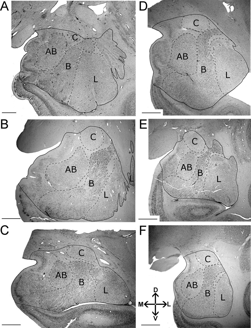
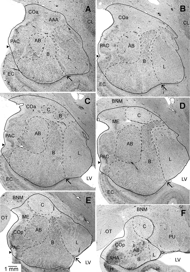
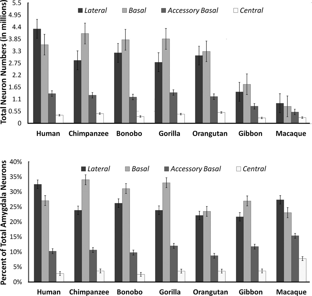
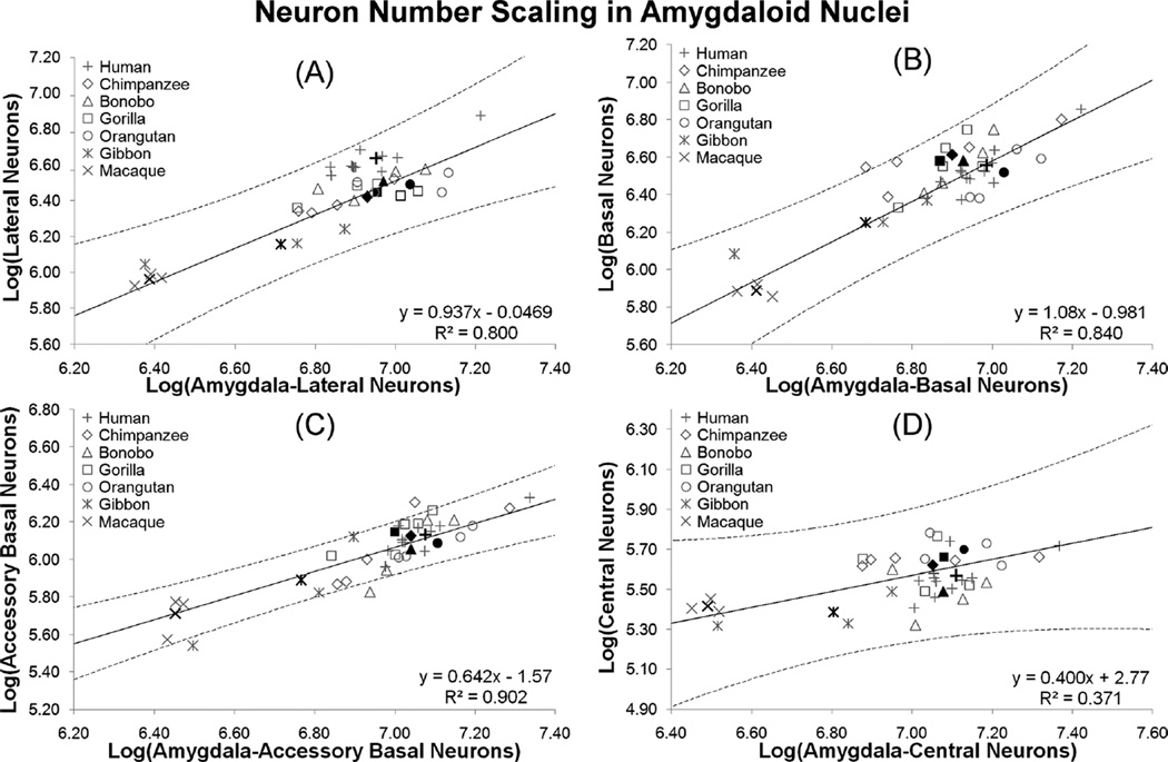
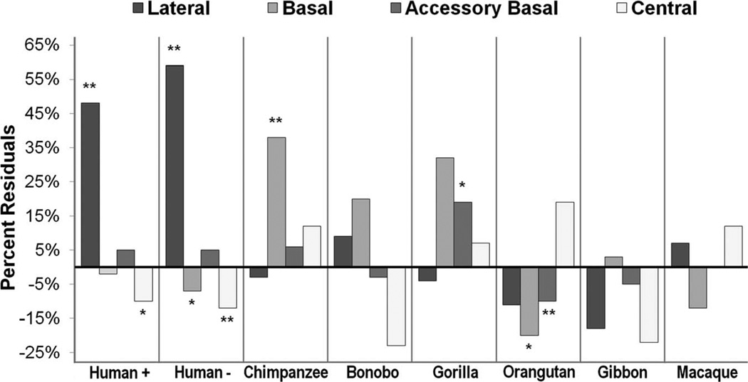
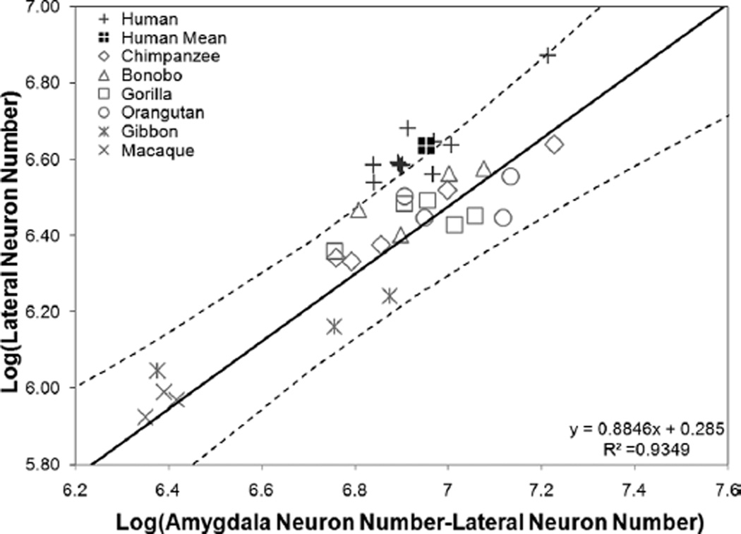
In human and nonhuman primates, the amygdala is known to play critical roles in emotional and social behavior. Anatomically, individual amygdaloid nuclei are connected with many neural systems that are either differentially expanded or conserved over the course of primate evolution. To address amygdala evolution in humans and our closest living relatives, the apes, we used design-based stereological methods to obtain neuron counts for the amygdala and each of four major amygdaloid nuclei (the lateral, basal, accessory basal, and central nuclei) in humans, all great ape species, lesser apes, and one monkey species. Our goal was to determine whether there were significant differences in the number or percent of neurons distributed to individual nuclei among species. Additionally, regression analyses were performed on independent contrast data to determine whether any individual species deviated from allometric trends. There were two major findings. In humans, the lateral nucleus contained the highest number of neurons in the amygdala, whereas in apes the basal nucleus contained the highest number of neurons. Additionally, the human lateral nucleus contained 59% more neurons than predicted by allometric regressions on nonhuman primate data. Based on the largest sample ever analyzed in a comparative study of the hominoid amygdala, our findings suggest that an emphasis on the lateral nucleus is the main characteristic of amygdala specialization over the course of human evolution.


Figures

Figure 1
Figure 1
Delineation of left amygdala and nuclei included in this analysis illustrating the consistency of borders across species. Images were taken from midrostrocaudal levels in the following primates: A: human. B: gorilla. C: orangutan. D: chimpanzee. E: gibbon. F: long tailed macaque. Abbreviations: AB, accessory basal nucleus; B, basal nucleus; C, central nucleus; L, lateral nucleus. Other amygdaloid nuclei are not represented in this comparative figure, but are highlighted in Figure 2. The human image (A) is modified from Schumann and Amaral (2005). Images follow radiological conventions. Scale bar = 2 mm in A–F.
Figure 2
Figure 2
A–F: A series of brightfield photomicrographs illustrating the boundaries of the amygdala, lateral, basal, accessory basal, and central nuclei in coronal sections of the left hemisphere of a chimpanzee. Images are from rostral (A,B), midrostrocaudal (C,D), and caudal (E,F) positions in the amygdala. Arrows point to the “notch” that separates the ventral borders of the lateral and basal nuclei. Small arrow-heads indicate the position of the semiannular sulcus used to mark the division between the cortical amygdaloid nuclei and the adjacent entorhinal cortex (anterior) or hippocampus (posterior). Abbreviations: AAA, anterior amygdaloid area; AB, accessory basal nucleus; AHA, amygdalohippocampal area; B, basal nucleus; BNM, basal nucleus of Meynert; C, central nucleus; CL, claustrum; COa, anterior cortical nucleus; COp, posterior cortical nucleus; EC, entorhinal cortex; H, hippocampus; I, intercalated nuclei; L, lateral nucleus; LV, lateral ventricle; ME, medial nucleus; OT, optic tract; PAC, periamygdaloid cortex; PU, putamen. Images follow radiological conventions. Scale bar = 1 mm in E (applies to A–F).
Figure 3
Figure 3
Tissue from the chimpanzee (A) lateral nucleus, (B) basal nucleus, and (C) central nucleus as viewed through a 100× objective, the magnification used for data collection. Morphological features of neurons (arrows) and glia (arrowheads) can be distinguished at this magnification. Scale bar = 15 µm in A–C.
Figure 4
Figure 4
Histograms indicating the average number of neurons (× 106) in the amygdala and four nuclei (top) and the average percent of total amygdala neurons distributed to the lateral, basal, and accessory basal nuclei across species (bottom) (n = 35). Error bars represent standard error.
Figure 5
Figure 5
Independent contrasts regression plotting the log of total amygdala neuron number against the log of the neuron numbers in (A) the lateral nucleus, (B) the basal nucleus, (C) the accessory basal nucleus, and (D) the central nucleus with all species included in each regression. Individual data points are plotted as open, gray markers and species mean values are plotted as closed, black markers.
Figure 6
Figure 6
Average percent residuals from regression equations in each nucleus for each species. Starred bars represent values that were statistically significant (**) or close to statistically significant (*) from a residual of 0. Human+, percent residual with all species included; Human−, percent residual excluding human data from the regression.
Figure 7
Figure 7
Independent contrasts regression plotting the log of amygdala neuron number against the log of neuron numbers in the lateral nucleus with humans excluded from the regression (n = 24).

References

    1. Adolphs R. Cognitive neuroscience of human social behaviour. Nat Rev Neurosci. 2003;4:165–178. - PubMed
    1. Adolphs R. What does the amygdala contribute to social cognition? Ann N Y Acad Sci. 2010;1191:42–61. - PMC - PubMed
    1. Allman JM, Tetreault NA, Hakeem AY, Manaye KF, Semendeferi K, Erwin JM, Park S, Goubert V, Hof PR. The von Economo neurons in frontoinsular and anterior cingulate cortex in great apes and humans. Brain Struct Funct. 2010;14:1–23. - PubMed
    1. Amaral D, Insausti R, Cowan WM. The entorhinal cortex of the monkey: I. Cytoarchitectonic organization. J Comp Neurol. 1987;264:326–355. - PubMed
    1. Amaral DG, Capitanio JP, Jourdain M, Mason WA, Mendoza SP, Prather M. The amygdala: is it an essential component of the neural network for social cognition? Neuropsychologia. 2003;41:235–240. - PubMed

Publication types