. 2012 Jul;80(7):2454-63.
doi: 10.1128/IAI.00018-12. Epub 2012 Apr 2.

Association of a protective monoclonal IgA with the O antigen of Salmonella enterica serovar Typhimurium impacts type 3 secretion and outer membrane integrity

Affiliations

Association of a protective monoclonal IgA with the O antigen of Salmonella enterica serovar Typhimurium impacts type 3 secretion and outer membrane integrity

Stephen J Forbes et al. Infect Immun. 2012 Jul.

Abstract

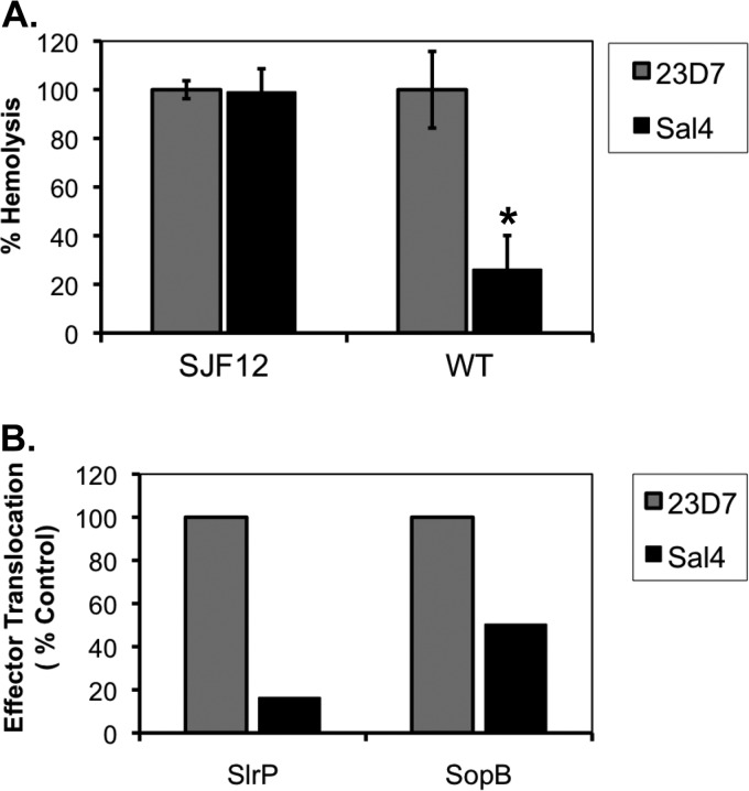
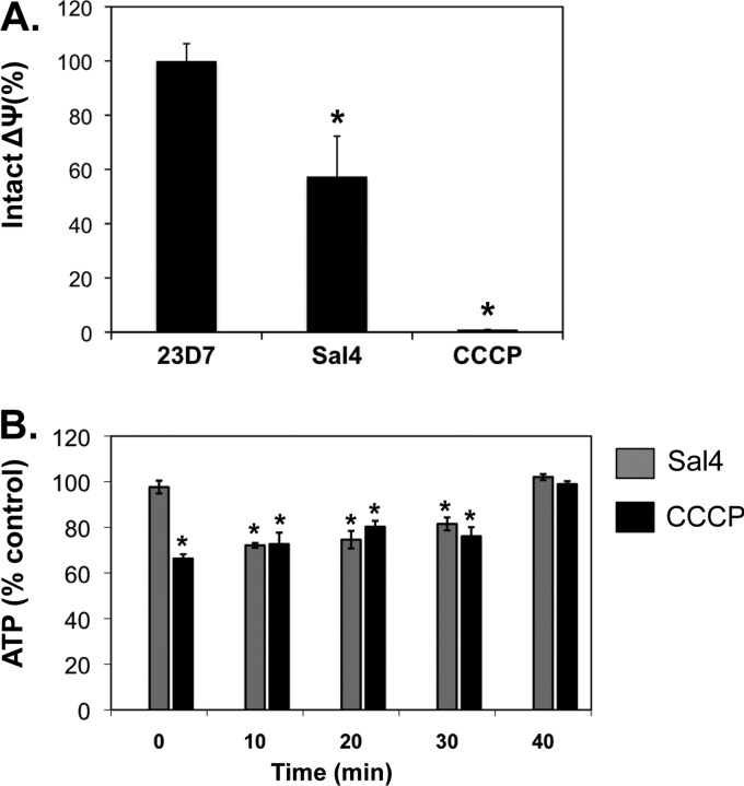
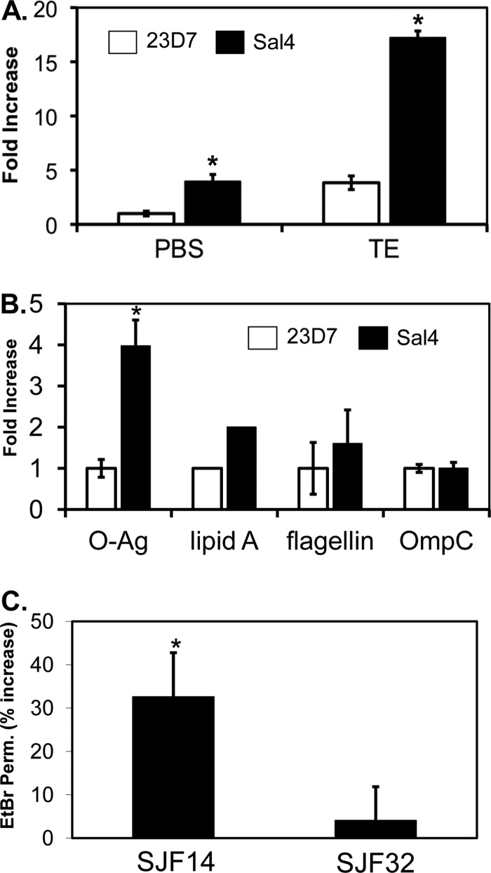
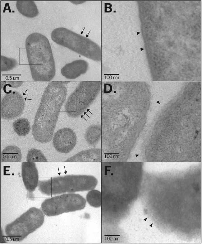
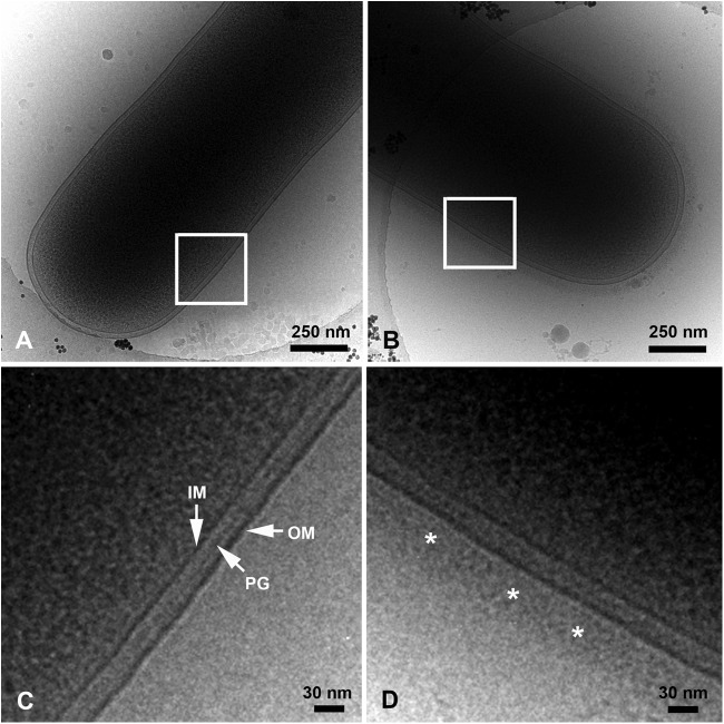
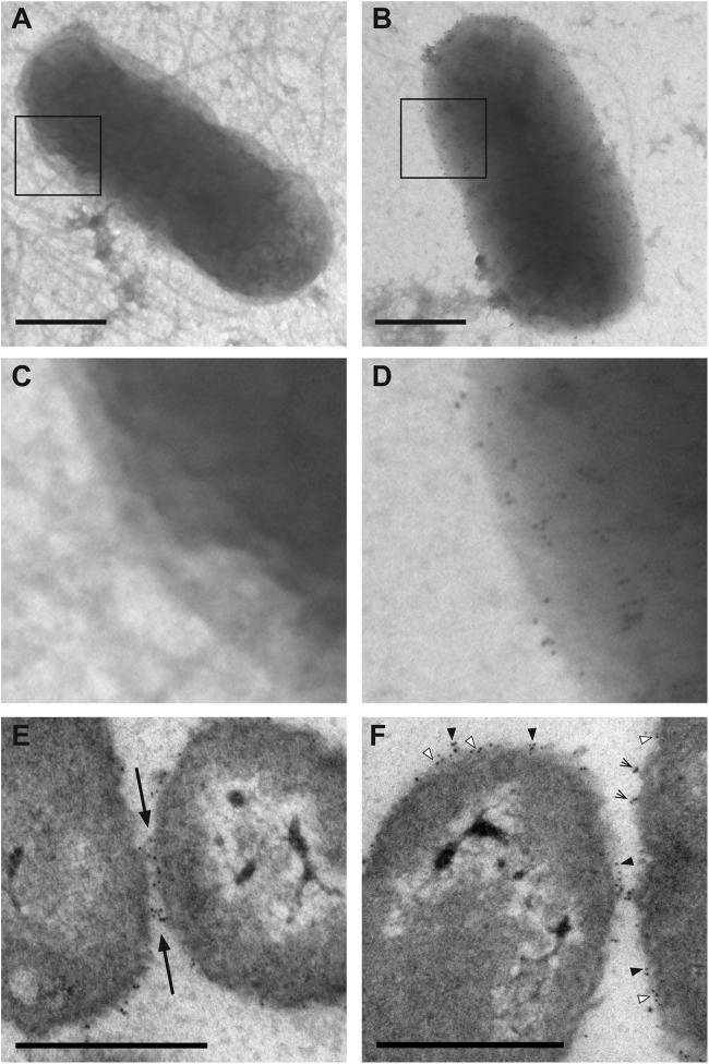
Invasion of intestinal epithelial cells by Salmonella enterica serovar Typhimurium is an energetically demanding process, involving the transfer of effector proteins from invading bacteria into host cells via a specialized organelle known as the Salmonella pathogenicity island 1 (SPI-1) type 3 secretion system (T3SS). By a mechanism that remains poorly understood, entry of S. Typhimurium into epithelial cells is inhibited by Sal4, a monoclonal, polymeric IgA antibody that binds an immunodominant epitope within the O-antigen (O-Ag) component of lipopolysaccharide. In this study, we investigated how the binding of Sal4 to the surface of S. Typhimurium influences T3SS activity, bacterial energetics, and outer membrane integrity. We found that Sal4 treatment impaired T3SS-mediated translocon formation and attenuated the delivery of tagged effector proteins into epithelial cells. Sal4 treatment coincided with a partial reduction in membrane energetics and intracellular ATP levels, possibly explaining the impairment in T3SS activity. Sal4's effects on bacterial secretion and energetics occurred concurrently with an increase in O-Ag levels in culture supernatants, alterations in outer membrane permeability, and changes in surface ultrastructure, as revealed by transmission electron microscopy and cryo-electron microscopy. We propose that Sal4, by virtue of its ability to bind and cross-link the O-Ag, induces a form of outer membrane stress that compromises the integrity of the S. Typhimurium cell envelope and temporarily renders the bacterium avirulent.


Figures

Fig 1
Fig 1
Reduced activity of the SPI-1 T3SS in the presence of Sal4. (A) Hemolysis as an indirect reporter of SPI-1 T3SS activity. Strains SJF12 and 14028 (WT) were incubated with Sal4 (5 μg/ml) or the ricin toxin-specific isotype control MAb 23D7 (5 μg/ml) for 15 min before the addition to 50% sheep whole blood, as described in Materials and Methods. SJF12 lacks the O5 epitope due to a chromosomal mutation in the oafA gene, which encodes a membrane-integral acetylase (58). The amount of hemolysis associated with each experimental condition was expressed as a percentage of the activity of the wild type or oafA strains alone. The data are the averages (with standard errors [SE]) from a single representative experiment done in triplicate. Sal4 treatment resulted in a statistically significant (*, P < 0.05) reduction in hemolytic activity, as determined by the Student t test. (B) Delivery of SPI-1 T3SS effector proteins into host cells. S. Typhimurium strains JS899 and JS898 expressing the chromosomally encoded effector protein SlrP or SopB, respectively, fused to GSK, were tested in HeLa cell invasion assays in the presence of Sal4 (5 μg/ml) or 23D7 (5 μg/ml), as described in Materials and Methods. Western blots of HeLa cell lysates probed with a GSK-specific antibody were subjected to densitometry. The values plotted on the y axis (“effector translocation”) are relative to the amount of GSK detected in HeLa cells infected with strain 14028 in the absence of either 23D7 or Sal4.
Fig 2
Fig 2
Reduced membrane potential and ATP levels in response to Sal4. (A) Mid-log-phase cells of S. Typhimurium were loaded with the dye JC-1 and then treated with Sal4 (5 μg/ml) or CCCP (100 μM) for 15 min. The samples were then visualized immediately by fluorescence microscopy, as described in Materials and Methods. The number of red (intact potential) and green (depleted potential) fluorescent bacteria in each sample was determined. The values obtained from the PBS-treated control cells were set to 100%. Each column represents the average value (with standard error) obtained from a minimum of three experiments. Significance, as determined using the Student t test, is indicated by asterisks: *, P < 0.05. (B) ATP levels in S. Typhimurium decline following treatment with Sal4. Mid-log-phase cultures of S. Typhimurium were diluted 1:10 into microtiter plates containing M9 minimal medium with Sal4 (5 μg/ml) or CCCP (100 μM) and incubated for 15 min before being assayed for ATP. Data are the percentage of ATP relative to wild-type S. Typhimurium treated with the isotype control antibody 23D7. Statistically significant differences (P < 0.05) between experimental and control samples, as determined by the Student t test, are indicated by asterisks.
Fig 3
Fig 3
Sal4 enhances the release of the O-Ag from the OM of S. Typhimurium. (A) Mid-log-phase cultures of S. Typhimurium were resuspended in PBS or T100E10 at pH 7.4 and then treated with 23D7 or Sal4 (5 μg/ml) for 15 min. Supernatants were then probed for O-Ag by dot blotting, as described in Materials and Methods. The membrane blots were analyzed by densitometry, and the data (fold increase) are presented relative to PBS control-treated cells. (B) Cell supernatants of S. Typhimurium treated with 23D7 or Sal4, as described for panel A, were subjected to immunoblotting with antibodies against O-Ag, flagellin, or OmpC. Lipid A levels in bacterial supernatants were detected using the LAL assay. Data are averages (with standard errors) from a single experiment done in triplicate. (C) Permeability of the bacterial OM to EtBr following Sal4 treatment. Mid-log-phase cultures of S. Typhimurium SJF14 (tolC::kan) or SJF32 (oafA126::Tn10d-Tc tolC::Kan) were treated with Sal4 (5 μg/ml) for 15 min and then incubated with EtBr (10 μg/ml) for 10 min. Cells were then subjected to quantitative fluorimetry. Baseline permeability values were obtained by measuring EtBr uptake in SJF14 and SJF32 treated with the isotype control antibody 23D7. The percent increase in permeability is defined as the amount of EtBr uptake in Sal4-treated cells divided by the amount obtained from 23D7-treated cells. Significance, as determined using the Student t test, is indicated by asterisks: *, P < 0.05.
Fig 4
Fig 4
Sal4 causes distortion of the bacterial envelope. S. Typhimurium strain 14028 was treated with PBS (A and B) or Sal4 (5 μg/ml) for 15 min (C and D) or 60 min (E and F) and then collected by centrifugation, fixed with glutaraldehyde, and processed for TEM, as described in Materials and Methods. The OM of control cells (arrows [A] or arrowheads [B]) were uniformly smooth. In contrast, cells treated with Sal4 for 15 min were surrounded by a fuzzy coat (arrows [C] or arrowheads [D]). The fuzzy coat was more pronounced after 60 min incubation with Sal4 (arrows [E] or arrowheads [F]). The boxed areas in panels A, C and E are enlarged in panels B, D, and F, respectively.
Fig 5
Fig 5
Cryo-electron micrographs of S. Typhimurium following treatment with Sal4. Control (PBS-treated [A and C]) or Sal4-treated (5 μg/ml for 15 min [B and D]) wild-type S. Typhimurium cells were spotted onto carbon-coated copper grids, plunge-frozen in liquid ethane, and imaged at −176°C using a JEM-4000FX electron microscope. Sal4-treated cells were surrounded by a fuzzy coat ∼50 to 60 nm thick (D, asterisks), which was absent from control cells. IM, inner membrane; PG, peptidoglycan layer; OM, outer membrane. The boxed areas in panels A and B are enlarged in panels C and D, respectively.
Fig 6
Fig 6
Association of Sal4 with the O-Ag of S. Typhimurium. (A to D) Mid-log-phase cultures of S. Typhimurium strain SJF12 (A and C) or 14028 (B and D) were spotted on Formvar-coated grids, fixed with glutaraldehyde, and then treated with Sal4-gold colloid (3 μg/ml) for 15 min. The grids were then visualized using a Zeiss 910 electron microscope, as described in Materials and Methods. As expected, Sal4-gold particles did not bind to the surface of SJF12 cells, which lack the O5 epitope due to a mutation in the oafA gene. In contrast, Sal4-gold particles were distributed uniformly around strain 14028 cells. The boxed areas in panels A and B are enlarged in panels C and D, respectively. (E and F) Mid-log-phase cultures of S. Typhimurium strain 14028 were treated with Sal4-gold colloid (5 μg/ml) for 15 min, collected by centrifugation, fixed with glutaraldehyde, and processed for TEM, as described in Materials and Methods. (E) Gold particles were often concentrated at sites of cell adhesion (Earrows). (F) Sal4-gold particles tended to be clustered on the surface (black arrowheads) but were occasionally embedded in the OM (white arrowheads) and situated at sites of membrane detachment (lined arrows). These observations suggest that Sal4 may both penetrate and promote the detachment of the LPS layer. Bars, 0.5 μm.

References

    1. Bakowski MA, et al. 2010. The phosphoinositide phosphatase SopB manipulates membrane surface charge and trafficking of the Salmonella-containing vacuole. Cell Host Microbe 7:453–462 - PubMed
    1. Becker LA, Bang IS, Crouch ML, Fang FC. 2005. Compensatory role of PspA, a member of the phage shock protein operon, in rpoE mutant Salmonella enterica serovar Typhimurium. Mol. Microbiol. 56:1004–1016 - PubMed
    1. Berg HC. 2003. The rotary motor of bacterial flagella. Annu. Rev. Biochem. 72:19–54 - PubMed
    1. Bernal-Bayard J, Ramos-Morales F. 2009. Salmonella type III secretion effector SlrP is an E3 ubiquitin ligase for mammalian thioredoxin. J. Biol. Chem. 284:27587–27595 - PMC - PubMed
    1. Blocker A, Komoriya K, Aizawa S. 2003. Type III secretion systems and bacterial flagella: insights into their function from structural similarities. Proc. Natl. Acad. Sci. U. S. A. 100:3027–3030 - PMC - PubMed

Publication types

LinkOut - more resources