. 2012 Jun 1;590(11):2739-50.
doi: 10.1113/jphysiol.2011.222935. Epub 2012 Apr 2.

The omega-6 fatty acid derivative 15-deoxy-Δ¹²,¹⁴-prostaglandin J2 is involved in neuroprotection by enteric glial cells against oxidative stress

Affiliations

The omega-6 fatty acid derivative 15-deoxy-Δ¹²,¹⁴-prostaglandin J2 is involved in neuroprotection by enteric glial cells against oxidative stress

Hind Abdo et al. J Physiol. .

Abstract

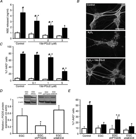
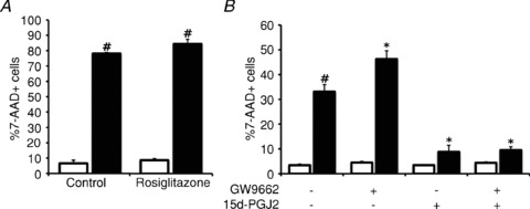
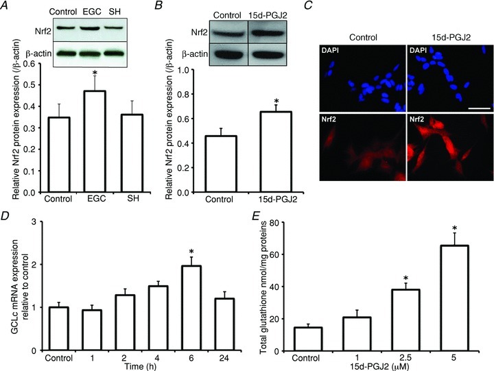
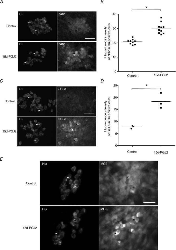
Increasing evidence suggests that enteric glial cells (EGCs) are critical for enteric neuron survival and functions. In particular, EGCs exert direct neuroprotective effects mediated in part by the release of glutathione. However, other glial factors such as those identified as regulating the intestinal epithelial barrier and in particular the omega-6 fatty acid derivative 15-deoxy-Δ¹²,¹⁴-prostaglandin J2 (15d-PGJ2) could also be involved in EGC-mediated neuroprotection. Therefore, our study aimed to assess the putative role of EGC-derived 15d-PGJ2 in their neuroprotective effects. We first showed that pretreatment of primary cultures of enteric nervous system(ENS)or humann euroblastoma cells (SH-SY5Y)with 15d-PGJ2 dose dependently prevented hydrogen peroxide neurotoxicity. Furthermore, neuroprotective effects of EGCs were significantly inhibited following genetic invalidation in EGCs of the key enzyme involved in 15d-PGJ2 synthesis, i.e. L-PGDS. We next showed that 15d-PGJ2 effects were mediated by an Nrf2 dependent pathway but were not blocked by PPARγ inhibitor (GW9662) in SH-SY5Y cells and enteric neurons. Finally, 15d-PGJ2 induced a significant increase in glutamate cysteine ligase expression and intracellular glutathione in SH cells and enteric neurons. In conclusion, we identified 15d-PGJ2 as a novel glial-derived molecule with neuroprotective effects in the ENS. This study further supports the concept that omega-6 derivatives such as 15d-PGJ2 might be used in preventive and/or therapeutic strategies for the treatment of enteric neuropathies.


Figures

Figure 1
Figure 1. EGC-derived 15d-PGJ2 (exerts neuroprotection against oxidative stress-induced cell death
A, primary cultures of ENS were treated or not treated with H2O2 (200 μm; 24 h; filled and open bars, respectively) with or without (control) previous treatments with 15d-PGJ2 (1, 2 or 3 μm; 72 h). NSE released in the medium was measured by immunoradiometric assay. Values are the mean ± SEM of from 6 to 11 independent experiments (#P < 0.05 as compared to cultures without H2O2; *P < 0.05 as compared to primary cultures treated with H2O2 but without 15d-PGJ2 pretreatment; two-way ANOVA followed by Bonferroni's post hoc test). B, primary cultures of ENS were treated or not treated with H2O2 (200 μm, 24 h), with or without previous treatment with 15d-PGJ2 (3 μm; 72 h). Immunostaining was performed with anti-β-tubulin III antibody. Images are representative of 4 independent experiments. Scale bar: 100 μm. C, SH-SY5Y cells were treated or not treated with H2O2 (200 μm; 24 h; filled and open bars, respectively), with or without (control) previous treatments with different concentrations of 15d-PGJ2 (0.1, 1 or 5 μm; 24 h). Neuronal cell death was analysed by measuring the cell permeability to 7-amino-actinomycin D (% 7-AAD+ cells) by flow cytometry. Values are the mean ± SEM from four independent experiments (#P < 0.05 as compared to conditions without H2O2; *P < 0.05 as compared to conditions treated with H2O2 but without 15d-PGJ2 pretreatment; two-way ANOVA followed by Bonferroni's post hoc test). D, EGCs were transfected with shRNA PTGDS (EGC shPTGDS) or with the shRNA PTGDS inefficient construction (EGC shMOCK). Inset: different non-transformed EGCs (NT; ROG) express L-PGDS. Immunoblot analysis using antibodies against change to L-PGDS or βactin were carried out from EGC extracts. Quantitative analysis was performed by measuring band densities with ImageJ. Values are the mean ± SEM from four independent experiments (*P < 0.05 as compared to EGCs or EGC shMOCK; one-way ANOVA followed by Tukey's post hoc test). E, SH-SY5Y cells were treated or not treated with H2O2 (200 μm; 24 h; filled and open bars, respectively) after 96 h co-culture with EGCs, EGC shPTGDS or EGC shMOCK. Neuronal cell death was analysed by measuring the cell permeability to 7-amino-actinomycin D (% 7-AAD+ cells) by flow cytometry. Values are the mean ± SEM from five independent experiments (#P < 0.05 as compared to control without H2O2, *P < 0.05 as compared to H2O2-treated SH-SY5Y cells alone; †P < 0.05 as compared to SH-SY5Y cells co-cultured with EGCs or EGC shMOCK; one-way ANOVA followed by Tukey's post hoc test).
Figure 2
Figure 2. 15d-PGJ2 neuroprotective effects are independent of the PPARγ pathway
A, SH-SY5Y cells were treated or not treated with H2O2 for (200 μm; 24 h; filled and open bars, respectively) after previous treatment with rosiglitazone (5 μm; 24 h). Neuronal cell death was analysed by measuring the cell permeability to 7-amino-actinomycin D (% 7-AAD+ cells) by flow cytometry. Values are the mean ± SEM from six independent experiments (#P < 0.05 as compared to untreated SH-SY5Y cells (control, open bar); one-way ANOVA followed by Tukey's post hoc test). B, SH-SY5Y cells were treated or not treated with H2O2 (200 μm; 24 h; filled and open bars, respectively) after treatment with 15d-PGJ2 (5 μm; 24 h), with or without 24 h pre-treatment with GW9662 (10 μm). Neuronal cell death was analysed by measuring the cell permeability to 7AAD (% 7AAD+ cells) by flow cytometry. Values are the mean ± SEM from four independent experiments (#P < 0.05 as compared to untreated conditions without H2O2 (open bar); *P < 0.05 as compared to untreated conditions with H2O2 (filled bar); one-way ANOVA followed by Tukey's post hoc test).
Figure 3
Figure 3. 15d-PGJ2 neuroprotective effects are associated with activation of the Nrf2 pathway
A, SH-SY5Y cells were cultured alone or co-cultured with EGCs or SH-SY5Y cells for 96 h. Immunoblot analysis using antibodies against Nrf2 or βactin was performed from SH-SY5Y extracts and was quantified by measuring band densities with ImageJ. Values are the mean ± SEM from seven independent experiments (*P < 0.05 as compared to control; one-way ANOVA followed by Tukey's post hoc test). B, SH-SY5Y cells were treated or not treated (control) with 15d-PGJ2 (5 μm; 5 h). Protein extracts were analysed for quantification with immunoblots using anti-Nrf2 and anti-βactin antibodies. Quantitative analysis was performed by measuring band densities with ImageJ. Values are the mean ± SEM of seven independent experiments (*P < 0.05 as compared to control; t test). C, SH-SY5Y cells were treated or not treated (control) with 15d-PGJ2 (5 μm; 5 h). Immunocytochemistry with anti-Nrf2 antibodies and DAPI labelling were performed and images are representative of three independent experiments. Scale bar: 100 μm. D, SH-SY5Y cells were treated or not (control) with 15d-PGJ2 (5 μm) for different time periods (1, 2, 4, 6 and 24 h). Quantification of GCLc mRNA was performed by RT–quantitative PCR as described in Methods. Values are the mean ± SEM of four independent experiments (*P < 0.05 as compared to control; one-way ANOVA followed by Tukey's post hoc test). E, SH-SY5Y cells were treated with varying concentrations of 15d-PGJ2 (1, 2.5 or 5 μm) for 24 h. Quantification of total glutathione was measured using enzymatic glutathione assay. Values are the mean ± SEM of four independent experiments (*P < 0.05 as compared to control; one-way ANOVA followed by Tukey's post hoc test).
Figure 4
Figure 4. 15d-PGJ2 neuroprotective effects on primary cultures of ENS are associated with Nrf2 pathway and glutathione synthesis
A, primary cultures of ENS were treated or not (control) with 15d-PGJ2 (3 μm, 2 h) and analysed by immunostaining with anti-Nrf2 or anti-Hu antibodies. Arrows represent examples of co-expression of Hu and Nrf2 proteins. Scale bar: 100 μm. B, quantitative analysis of Nrf2 protein expression in enteric neurons. Mean fluorescence intensity of Nrf2 corresponding to each enteric neuron (Hu-positive cell) was determined from ImageJ software analysis of control ENS cultures (9 images, 19–49 enteric neurons per image, two independent experiments, 305 enteric neurons analysed) and after 2 h of 15d-PGJ2-treatment (10 images, 21–39 enteric neurons per image, two independent experiments, 292 enteric neurons analysed). Mean fluorescence intensity was corrected by measuring background for each image. Each symbol represents the mean value of fluorescence intensity in enteric neurons per analysed image (*P < 0.001 as compared to control; t test). C, primary cultures of ENS were treated or not treated (control) with 15d-PGJ2 (3 μm; 24 h) and analysed by immunostaining with anti-GCLc or anti-Hu antibodies. Arrows represent examples of co-expression of Hu and GCLc proteins. Scale bar: 100 μm. D, quantitative analysis of GCLc protein expression in enteric neurons. Mean fluorescence intensity of GCLc corresponding to each enteric neuron (Hu-positive cell) was determined from ImageJ software analysis of control ENS cultures (3 images, 24–37 enteric neurons per image, 97 enteric neurons analysed) and after 2 h of 15d-PGJ2-treatment (3 images, 17–27 enteric neurons per image, 69 enteric neurons analysed). Mean fluorescence intensity was corrected by measuring background for each image. Each symbol represents the mean value per analysed image of fluorescence intensity in enteric neurons (*P < 0.01 as compared to control; t test). E, primary cultures of ENS were treated or not treated (control) with 15d-PGJ2 (3 μm; 24 h). Following treatment, monochlorobimane (MCB, 60 μm) was directly added to the medium and incubated for 20 min at 37°C before fixation and standard immunostaining procedures with anti-Hu antibodies. Treatment of primary cultures of ENS with 3 μm 15d-PGJ2 for 24 h induced an increase of total glutathione conjugated to MCB in enteric neurons (Hu-positive cells) as compared to untreated cultures (Control). Arrows represent examples of co-expression of Hu and conjugated MCB. Scale bar: 100 μm.

Comment in

  • Enteric neuroprotection.
    Luyer MD. Luyer MD. J Physiol. 2012 Jun 15;590(12):2827. doi: 10.1113/jphysiol.2012.234534. J Physiol. 2012. PMID: 22707588 Free PMC article. No abstract available.

Similar articles

Cited by

References

    1. Abdo H, Derkinderen P, Gomes P, Chevalier J, Aubert P, Masson D, Galmiche JP, Vanden Berghe P, Neunlist M, Lardeux B. Enteric glial cells protect neurons from oxidative stress in part via reduced glutathione. FASEB J. 2010;24:1082–1094. - PubMed
    1. Aoun P, Watson DG, Simpkins JW. Neuroprotective effects of PPARγ agonists against oxidative insults in HT-22 cells. Eur J Pharmacol. 2003;472:65–71. - PubMed
    1. Aube AC, Cabarrocas J, Bauer J, Philippe D, Aubert P, Doulay F, Liblau R, Galmiche JP, Neunlist M. Changes in enteric neurone phenotype and intestinal functions in a transgenic mouse model of enteric glia disruption. Gut. 2006;55:630–637. - PMC - PubMed
    1. Bach-Ngohou K, Mahé MM, Aubert P, Abdo H, Boni S, Bourreille A, Denis MG, Lardeux B, Neunlist M, Masson D. Enteric glia modulate epithelial cell proliferation and differentiation through 15-deoxy-Δ12,14-prostaglandin J2. J Physiol. 2010;588:2533–2544. - PMC - PubMed
    1. Bush TG, Savidge TC, Freeman TC, Cox HJ, Campbell EA, Mucke L, Johnson MH, Sofroniew MV. Fulminant jejuno-ileitis following ablation of enteric glia in adult transgenic mice. Cell. 1998;93:189–201. - PubMed

Publication types

LinkOut - more resources