. 2012 Jul 1;590(13):3185-202.
doi: 10.1113/jphysiol.2011.224873. Epub 2012 Apr 2.

The chemokine CXCL12 and the HIV-1 envelope protein gp120 regulate spontaneous activity of Cajal-Retzius cells in opposite directions

Affiliations

The chemokine CXCL12 and the HIV-1 envelope protein gp120 regulate spontaneous activity of Cajal-Retzius cells in opposite directions

Ivan Marchionni et al. J Physiol. .

Abstract

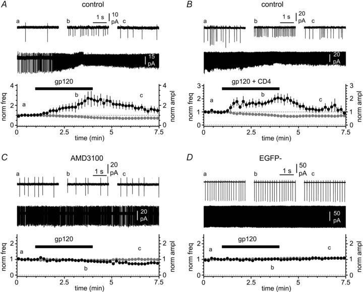
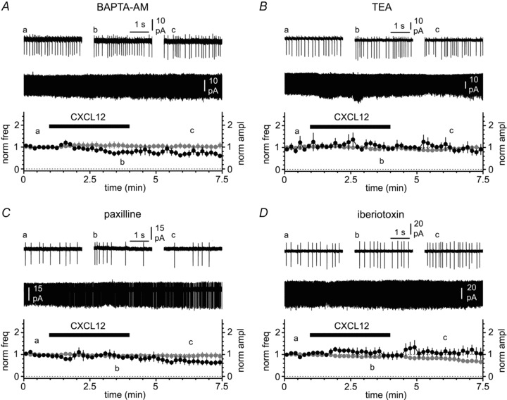
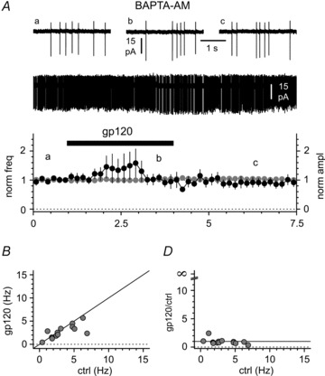
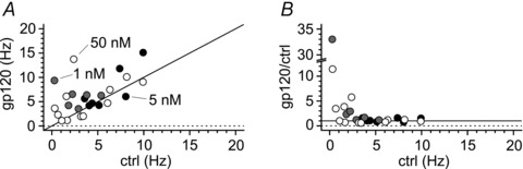
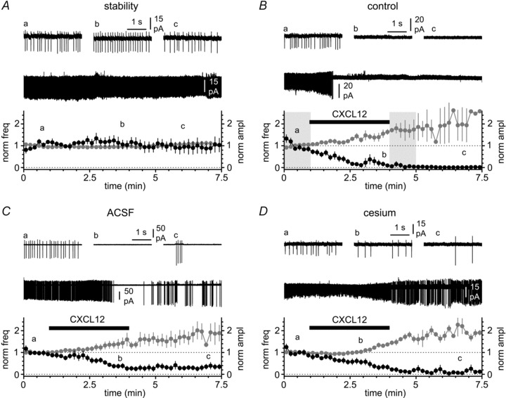
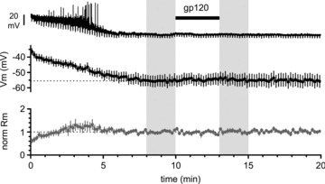
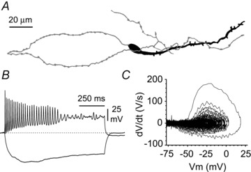
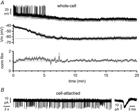
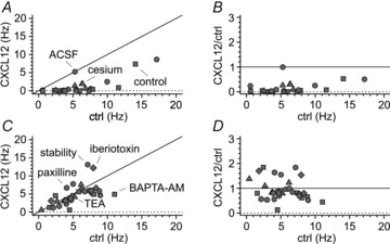
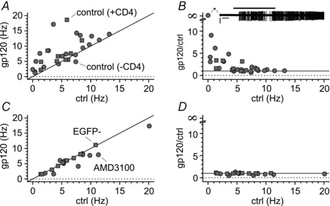
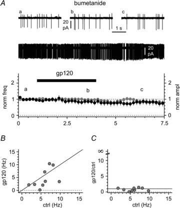
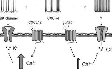
Activation of the CXC chemokine receptor 4 (CXCR4) in Cajal–Retzius cells by CXC chemokine ligand 12 (CXCL12) is important for controlling their excitability. CXCR4 is also a co-receptor for the glycoprotein 120 (gp120) of the envelope of the human immunodeficiency virus type 1 (HIV-1), and binding of gp120 to CXCR4 may produce pathological effects. In order to study CXCR4-dependent modulation of membrane excitability, we recorded in cell-attached configuration spontaneous action currents from hippocampal stratum lacunosum-moleculare Cajal–Retzius cells of the CXCR4-EGFP mouse. CXCL12 (50 nM) powerfully inhibited firing independently of synaptic transmission, suggesting that CXCR4 regulates an intrinsic conductance. This effect was prevented by conditioning slices with BAPTA-AM (200 μM), and by blockers of the BK calcium-dependent potassium channels (TEA (1 mM), paxilline (10 μM) and iberiotoxin (100 nM)). In contrast, exposure to gp120 (pico- to nanomolar range, alone or in combination with soluble cluster of differentiation 4 (CD4)), enhanced spontaneous firing frequency. This effect was prevented by the CXCR4 antagonist AMD3100 (1 μM) and was absent in EGFP-negative stratum lacunosum-moleculare interneurons. Increased excitability was prevented by treating slices with BAPTA-AM or bumetanide, suggesting that gp120 activates a mechanism that is both calcium- and chloride-dependent. In conclusion, our results demonstrate that CXCL12 and gp120 modulate the excitability of Cajal–Retzius cells in opposite directions. We propose that CXCL12 and gp120 either generate calcium responses of different strength or activate distinct pools of intracellular calcium, leading to agonist-specific responses, mediated by BK channels in the case of CXCL12, and by a chloride-dependent mechanism in the case of gp120.


Figures

Figure 6
Figure 6. gp120 increases the excitability of Cajal–Retzius cells via the chemokine receptor CXCR4 in an apparent CD4-independent fashion
A, application of gp120 (black bar, 200 pm) increases spike frequency and decreases the amplitude of the action currents. Insets illustrate the entire recording and selected portions before the application of gp120 (a), in its presence (b) and after its removal (c). B, as in A, but gp120 was primed with equimolar CD4. Notice the similarity of the effects on frequency and amplitude compared to gp120 alone in A. C, as in A and B, but in the presence of the CXCR4 antagonist AMD3100 (1 μm). Notice that the antagonist prevents the gp120-induced modulation. D, as in the previous panels, but recordings were made from stratum EGFP non-expressing neurons in stratum lacunosum-moleculare. Notice the absence of any effect induced by gp120.
Figure 4
Figure 4. Modulation of spontaneous firing by CXCL12 is mediated by BK-type calcium-activated potassium channels
A, lack of effects on frequency and amplitude of action currents when CXCL12 is applied (black bar, 50 nm) to slices pre-conditioned by BAPTA-AM. Insets illustrate the entire recording and selected portions before the application of CXCL12 (a), in its presence (b) and after its removal (c). B, C and D, as in A, but in the constant presence of TEA (1 mm), paxilline (10 μm) and iberiotoxin (100 nm), respectively. Notice that all these blockers of BK channels prevent the effect of CXCL12.
Figure 9
Figure 9. gp120-induced modulation of activity in Cajal–Retzius cells depends on intracellular calcium
A, application of gp120 (200 pm, black bar) to slices conditioned by BAPTA-AM does not change either spontaneous firing frequency or action current amplitude. Insets illustrate the entire recording and selected portions before the application of gp120 (a), in its presence (b) and after its removal (c). B, the lack of effect in BAPTA-AM-treated slices is not due to frequency-dependent effects. The summary plot of the individual results shows that for this experiment we pre-selected cells with low control firing rates. Notice the clustering of the data around the identity line (black line). C, the lack of effect is apparent in a frequency range that is associated with increased excitability in non-conditioned slices (compare this panel with Fig. 7B).
Figure 11
Figure 11. The increase in intrinsic firing of Cajal–Retzius cells is maintained when gp120 is applied in the nanomolar range (1–50 nm)
A, plot of results from individual experiments where gp120 was applied at different nanomolar concentrations (1 nm, grey circles, 5 nm, black circles, 50 nm, white circles). Notice the scatter of the data above the identity line (black line). B, normalized effect of gp120 application on spontaneous frequencies under the same experimental conditions as in A. Notice that, similarly to what is shown in Fig. 7B, the effect of gp120 is much stronger when tested on cells with low control frequencies.
Figure 3
Figure 3. Modulation of spontaneous firing by CXCL12 application in cell-attached conditions
A, spontaneous firing can be monitored by measuring the frequency (black circles) and amplitude (grey circles) of the action currents. Notice that in the absence of CXCL12 application both parameters remain stable in control solution (ACSF + synaptic blockers). Insets show the entire recording and selected portions at different time points (a, b and c). B, application of CXCL12 (black bar, 50 nm) dramatically decreases the frequency of the action currents and concomitantly increases their amplitude. Insets illustrate the entire recording and selected portions before the application of CXCL12 (a), in its presence (b) and after its removal (c). Notice that the effect is long-lasting. The shaded areas indicate the time windows used for the quantification of firing frequency and action current amplitude in control and after modulation by the chemokine (see Methods). C, as in B, but experiments were performed in the absence of synaptic blockers. Notice the persistence of a strong effect both on frequency and amplitude. D, as in A and B, but in the constant presence of extracellular cesium (2 mm), to test the involvement of potassium inward rectifiers in the response produced by CXCL12. Notice the persistence of the effect.
Figure 8
Figure 8. The effects of gp120 are lost when whole-cell recording conditions are used
Top panel: original recording from a Cajal–Retzius cell, which progressively hyperpolarizes its membrane potential following breakthrough (beginning of the trace, t= 0 min). Downward deflections are the responses to regularly injected hyperpolarizing current pulses to monitor the membrane input resistance (−5 pA, 1.5 s duration). Middle panel: summary plot from several experiments showing a total lack of effect of gp120 application (200 pm, black bar). Lower panel: summary graph of the estimated membrane input resistance. The shaded areas indicate the time windows used for the quantification of membrane potential and cell input resistance in control and after application of gp120.
Figure 1
Figure 1. Basic characteristics of hippocampal Cajal–Retzius cells of the CXCR4-EGFP mouse
A, stereotypical appearance of an EGFP-expressing Cajal–Retzius cell of CA1 stratum lacunosum-moleculare (Marchionni et al. 2010). Notice the tadpole-like morphology (soma/dendrite: black, axon: grey). B, basic electrophysiological properties of Cajal–Retzius cells. Notice the rapidly decreasing amplitude of the spikes with membrane depolarization and the sag elicited by hyperpolarizing current injections (pulses were +60 pA and −25 pA, respectively, 1 s duration). C, phase plot of the record shown in B illustrating the rapidly decreasing dV/dt at more depolarizing potentials.
Figure 2
Figure 2. Cajal–Retzius cells are particularly sensitive to whole-cell recording conditions
A, top panel: a spontaneously firing Cajal–Retzius cell loses its spontaneous activity after breakthrough (beginning of the trace, t= 0 min) and acquires a membrane resting potential. Downward deflections are the responses to regularly injected hyperpolarizing current pulses to monitor the membrane input resistance (−5 pA, 1.5 s duration). Middle panel: summary plot from several experiments showing a robust hyperpolarization occurring within the first ∼5 min from breakthrough. Lower panel: summary graph of the estimated membrane input resistance (Rm). B, spontaneous firing monitored in cell-attached recording configuration. Notice the presence of action currents shown at slower (left panel) and faster (right panel) time scales.
Figure 5
Figure 5. Plots of individual results from several CXCL12 experiments and stability test
A, summary graph illustrating frequencies of spontaneous firing before (ctrl) and after the application of CXCL12 (CXCL12). Notice that application of the chemokine in control solution (squares), ACSF (circles) or in the constant presence of cesium (triangles) results in almost all the experiments in frequencies below the identity line (black line). B, normalized effect of CXCL12 application on spontaneous frequencies under the same experimental conditions as in A. Notice that frequency is strongly decreased irrespective of the frequencies measured before CXCL12 application. C and D, as in A and B, respectively, but for experiments in which no modulation of firing is present (stability: circles, BAPTA-AM: squares, TEA: triangles, paxilline: hexagons, iberiotoxin: diamonds). Notice the individual data points in C clustering around the identity line, and the lack of normalized effects in D.
Figure 7
Figure 7. The increased excitability caused by gp120 appears to be frequency dependent
A, plot of results from individual experiments (gp120 alone: control, grey circles; gp120 primed and in the constant presence of CD4: CD4, grey squares). Notice the scatter of the data above the identity line (black line). B, normalized effect of gp120 application on spontaneous frequencies under the same experimental conditions as in A. Notice that the effect of gp120 is much stronger when tested on cells with low control frequencies. The inset (asterisk) shows that gp120 application could trigger firing in a resting Cajal–Retzius cell. C and D, as in A and B, respectively, but for experiments where the effect of gp120 was either blocked by the CXCR4 antagonist AMD3100 (grey circles) or absent (EGFP non-expressing cells: EGFP, grey squares). Notice the clustering of the data around the identity line and the lack of effects independent of the control frequency.
Figure 10
Figure 10. Effect of the NKCC1 blocker bumetanide (50 μm) on gp120-induced modulation of activity in Cajal–Retzius cells
A, application of gp120 (200 pm, black bar) to slices pre-incubated and in the constant presence of bumetanide does not change either spontaneous firing frequency or action current amplitude. Insets illustrate the entire recording and selected portions before the application of gp120 (a), in its presence (b) and after its removal (c). B, the lack of effect in bumetanide-treated slices is not due to frequency-dependent effects. The summary plot of the individual results shows that for this experiment we pre-selected cells with low control firing rates. Notice also that in some individual cells gp120 appeared to actually decrease firing frequency (black line = identity line). C, the lack of effect is apparent in a frequency range that is associated with increased excitability in non-conditioned slices (compare this panel with Fig. 7B).
Figure 12
Figure 12. Schematic representation of the proposed hypothesis of biased agonism at the CXCR4 receptor in Cajal–Retzius cells
Activation of CXCR4 by different agonists (CXCL12 vs. gp120) leads to quantitatively different intracellular calcium responses (larger vs. smaller arrow), which activate specific conductances: BK channels in the case of CXCL12 and a chloride-dependent mechanism in the case of gp120 (here proposed as a calcium-dependent chloride channel, indicated by the question mark). While opening of a potassium conductance leads to reduced excitability, the chloride mechanism modulated by gp120 increases Cajal–Retzius cell spontaneous firing.

Comment in

Similar articles

Cited by

References

    1. Abraham H, Meyer G. Reelin-expressing neurons in the postnatal and adult human hippocampal formation. Hippocampus. 2003;13:715–727. - PubMed
    1. Abraham H, Toth Z, Seress L. A novel population of calretinin positive neurons comprises reelin-positive Cajal–Retzius cells in the hippocampal formation of the adult domestic pig. Hippocampus. 2004;14:385–401. - PubMed
    1. Achilles K, Okabe A, Ikeda M, Shimizu-Okabe C, Yamada J, Fukuda A, Luhmann HJ, Kilb W. Kinetic properties of Cl uptake mediated by Na+-dependent K+-2Cl cotransport in immature rat neocortical neurons. J Neurosci. 2007;27:8616–8627. - PMC - PubMed
    1. Aguiló A, Schwartz TH, Kumar VS, Peterlin ZA, Tsiola A, Soriano E, Yuste R. Involvement of Cajal–Retzius neurons in spontaneous correlated activity of embryonic and postnatal layer 1 from wild-type and reeler mice. J Neurosci. 1999;19:10856–10868. - PMC - PubMed
    1. Ahuja S, Smith SO. Multiple switches in G protein-coupled receptor activation. Trends Pharmacol Sci. 2009;30:494–502. - PubMed

Publication types

LinkOut - more resources