

A myelin gene causative of a catatonia-depression syndrome upon aging
- PMID: 22473874
- PMCID: PMC3443947
- DOI: 10.1002/emmm.201200230
A myelin gene causative of a catatonia-depression syndrome upon aging
Abstract
Severe mental illnesses have been linked to white matter abnormalities, documented by postmortem studies. However, cause and effect have remained difficult to distinguish. CNP (2',3'-cyclic nucleotide 3'-phosphodiesterase) is among the oligodendrocyte/myelin-associated genes most robustly reduced on mRNA and protein level in brains of schizophrenic, bipolar or major depressive patients. This suggests that CNP reduction might be critical for a more general disease process and not restricted to a single diagnostic category. We show here that reduced expression of CNP is the primary cause of a distinct behavioural phenotype, seen only upon aging as an additional 'pro-inflammatory hit'. This phenotype is strikingly similar in Cnp heterozygous mice and patients with mental disease carrying the AA genotype at CNP SNP rs2070106. The characteristic features in both species with their partial CNP 'loss-of-function' genotype are best described as 'catatonia-depression' syndrome. As a consequence of perturbed CNP expression, mice show secondary low-grade inflammation/neurodegeneration. Analogously, in man, diffusion tensor imaging points to axonal loss in the frontal corpus callosum. To conclude, subtle white matter abnormalities inducing neurodegenerative changes can cause/amplify psychiatric diseases.
Copyright © 2012 EMBO Molecular Medicine.
Figures
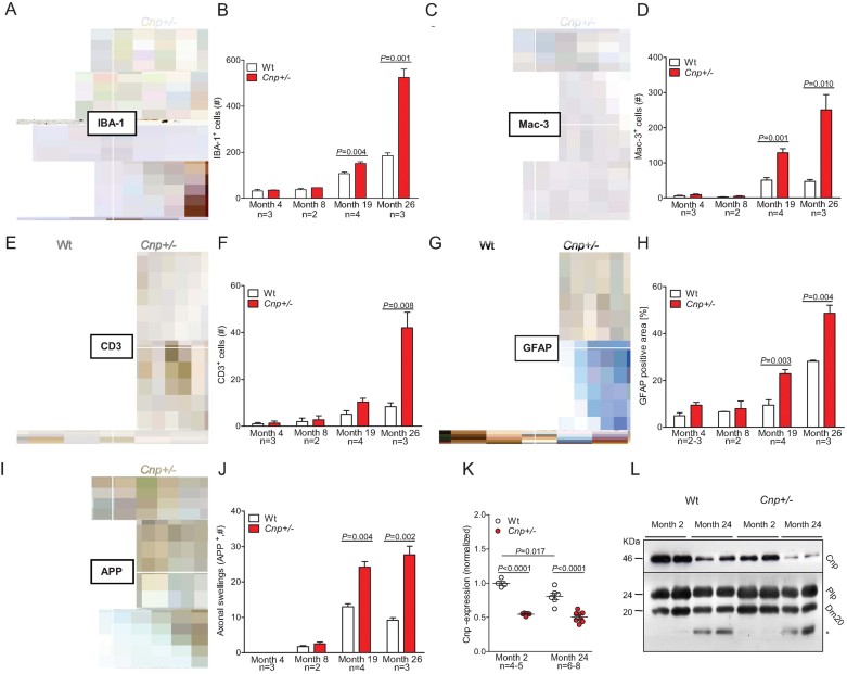
Representative microscopic images of the corpus callosum from 4 months (upper panels) and 26 months (lower panels) old Wt and Cnp+/− mice, immunostained for IBA-1; scale bar 20 µm.
Bar graph gives the age-dependent quantification of the total number of IBA-1 positive microglia in the corpus callosum of Wt and Cnp+/− mice. For all quantifications (B, D, F, H, J), n numbers indicated; mean ± s.e.m. presented; two-sided Student's t-test used.
Representative microscopic images of the corpus callosum from 4 months (upper panels) and 26 months (lower panels) old Wt and Cnp+/− mice, immunostained for Mac-3; scale bar 20 µm.
Bar graph gives the age-dependent quantification of the total number of Mac-3 positive microglia in the corpus callosum of Wt and Cnp+/− mice.
Representative microscopic images of the corpus callosum from 4 months (upper panels) and 26 months (lower panels) old Wt and Cnp+/− mice, immunostained for CD3; black arrows exemplify respective positive cells; scale bar 20 µm.
Bar graph gives the age-dependent quantification of the total number of CD3 positive T-lymphocytes in the corpus callosum, striatum and anterior commissure of Wt and Cnp+/− mice.
Representative microscopic images of the corpus callosum from 4 months (upper panels) and 26 months (lower panels) old Wt and Cnp+/− mice, immunostained for GFAP; scale bar 20 µm.
Densitometrical quantification of the GFAP positive area in the corpus callosum.
Representative microscopic images of the striatum from 4 months (upper panels) and 26 months (lower panels) old Wt and Cnp+/− mice, immunostained for APP; black arrows exemplify respective positive cells; scale bar 20 µm.
Bar graph gives the age-dependent quantification of the APP positive axonal swellings in the corpus callosum, striatum and anterior commissure of Wt and Cnp+/− mice.
Cnp mRNA expression level of Wt and Cnp+/− mice at months 2 and 24, normalized to mean value of ATP synthase subunit beta (Atp5b) and acidic ribosomal phosphoprotein P0 (Rplp0) as housekeeper genes and to 2 months old Wt (1.0); mean ± s.e.m. presented; two-sided Student's t-test used.
Cnp protein expression of Wt and Cnp+/− mice at months 2 and 24, compared to Plp as control protein of compact myelin; * low-size band detected in aged brain myelin with the Plp antibody directed against the C-terminus of PLP/DM20.
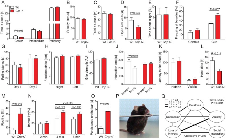
A-C Open arm parameters.
D. Elevated plus maze.
E. Light/dark box paradigm.
F. Baseline freezing in the context and cue memory task of fear conditioning.
G. Rota-rod.
H. Gait analysis.
I. Grip strength.
J. Sociability testing in the three-partite chamber.
K. Buried-food finding test – latency to find hidden versus visible food pellets.
L. Hole board.
M. Floating rate in a 90 s swim trial.
N. Tail suspension test.
O. Bar test for catatonia.
P. Typical posture of a catatonic Cnp+/− mouse during the bar test; see also videos of Supporting Information.
Q. Behavioural composite score displayed as intercorrelation network of Z-transformed items. Line thickness indicates the degree of correlation between 2 respective items. The composite score differs between genotypes (p = 0.0001). For all behavioural experiments, 24 months old mice were used: Wt n = 9–11 and Cnp+/− n = 10–16; mean ± s.e.m. presented; two-sided or paired t-tests used where applicable.
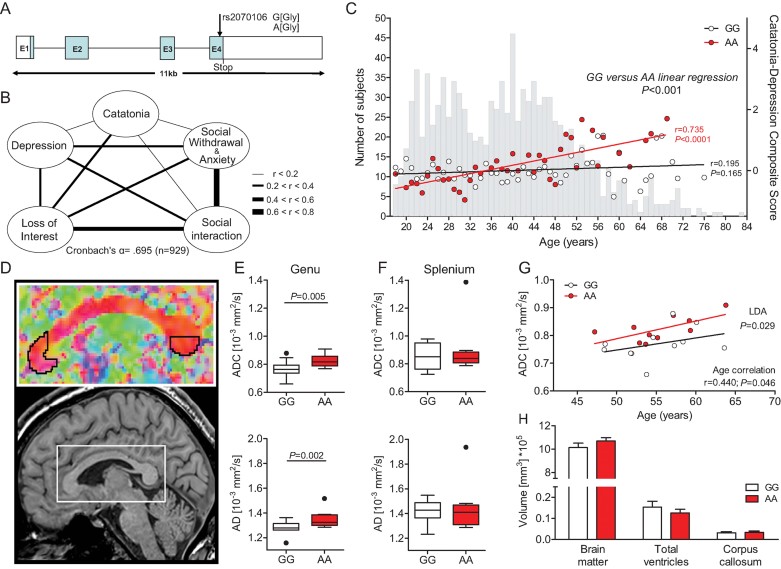
A. Schematic view of the human CNP gene structure and location of the synonymous SNP rs2070106 (A/G).
B. Intercorrelation network of all Z-transformed items of the catatonia-depression composite in the GRAS population. Line thickness indicates the degree of correlation between two respective items.
C. Correlation of genotypes with the catatonia-depression composite score across age groups. Grey bars in the background display the age distribution of the total GRAS sample of schizophrenic patients (n = 1048). Red or white circles denote mean values of the composite score for the respective age group and genotype (red, AA; black, GG). Linear regression lines of the genotypes dissociate after the age of 40 years. Pearson product-moment correlation applied.
D. Diffusion tensor imaging (DTI) study selecting the frontal (genu) and caudal (splenium) areas of the corpus callosum as regions of interest.
E,F ADC and AD values plotted according to rs2070106 homozygosity status in genu (E, target region) and splenium (F, control region) of the corpus callosum in a subgroup of schizophrenic individuals >40 years of age (GG n = 11 and AA n = 10); results corrected for chlorpromazine equivalents (CPZ). Mean ± s.e.m. presented and ANCOVA applied.
G. Correlation of ADC and age in AA and GG genotypes; linear discriminant analysis (LDA) with genotype as grouping variable and ADC and age as independent variables. Pearson product-moment correlation applied.
H. Magnetic resonance imaging (MRI) volumetric comparison of brain matter, ventricular system and corpus callosum between genotypes. Mean ± s.e.m. presented; two-sided Student's t-test applied.
Similar articles
-
Microglia ablation alleviates myelin-associated catatonic signs in mice.J Clin Invest. 2018 Feb 1;128(2):734-745. doi: 10.1172/JCI97032. Epub 2017 Dec 18. J Clin Invest. 2018. PMID: 29252214 Free PMC article.
-
Micro(glial)-managing executive function: white matter inflammation drives catatonia.J Clin Invest. 2018 Feb 1;128(2):564-566. doi: 10.1172/JCI98761. Epub 2017 Dec 18. J Clin Invest. 2018. PMID: 29252213 Free PMC article.
-
Cellular and subcellular distribution of 2',3'-cyclic nucleotide 3'-phosphodiesterase and its mRNA in the rat central nervous system.J Neurochem. 1988 Sep;51(3):859-68. doi: 10.1111/j.1471-4159.1988.tb01822.x. J Neurochem. 1988. PMID: 2842456
-
[Catatonia or depression: the GC rat strain as an animal model of psychopathology].Genetika. 2004 Jun;40(6):827-34. Genetika. 2004. PMID: 15341273 Review. Russian.
-
Brain imaging in catatonia: systematic review and directions for future research.Psychol Med. 2020 Jul;50(10):1585-1597. doi: 10.1017/S0033291720001853. Epub 2020 Jun 16. Psychol Med. 2020. PMID: 32539902
Cited by
-
Phenotype-Based Genetic Association Studies (PGAS)-Towards Understanding the Contribution of Common Genetic Variants to Schizophrenia Subphenotypes.Genes (Basel). 2014 Feb 27;5(1):97-105. doi: 10.3390/genes5010097. Genes (Basel). 2014. PMID: 24705289 Free PMC article.
-
Determinants of ligand binding and catalytic activity in the myelin enzyme 2',3'-cyclic nucleotide 3'-phosphodiesterase.Sci Rep. 2015 Nov 13;5:16520. doi: 10.1038/srep16520. Sci Rep. 2015. PMID: 26563764 Free PMC article.
-
Nanobodies against the myelin enzyme CNPase as tools for structural and functional studies.J Neurochem. 2025 Jan;169(1):e16274. doi: 10.1111/jnc.16274. J Neurochem. 2025. PMID: 39655780 Free PMC article.
-
Cell-autonomous requirement of TDP-43, an ALS/FTD signature protein, for oligodendrocyte survival and myelination.Proc Natl Acad Sci U S A. 2018 Nov 13;115(46):E10941-E10950. doi: 10.1073/pnas.1809821115. Epub 2018 Oct 29. Proc Natl Acad Sci U S A. 2018. PMID: 30373824 Free PMC article.
-
Interaction between Oligodendrocytes and Interneurons in Brain Development and Related Neuropsychiatric Disorders.Int J Mol Sci. 2024 Mar 23;25(7):3620. doi: 10.3390/ijms25073620. Int J Mol Sci. 2024. PMID: 38612430 Free PMC article. Review.
References
-
- Amir S. Catalepsy induced by body pinch: relation to stress-induced analgesia. Ann N Y Acad Sci. 1986;467:226–237. - PubMed
-
- APA. Diagnostic and Statistical Manual of Mental Disorders: DSM-IV-TR. Washington: American Psychiatric Association; 2000.
-
- Arora M, Praharaj SK. Butterfly glioma of corpus callosum presenting as catatonia. World J Biol Psychiatry. 2007;8:54–55. - PubMed
-
- Aston C, Jiang L, Sokolov BP. Transcriptional profiling reveals evidence for signaling and oligodendroglial abnormalities in the temporal cortex from patients with major depressive disorder. Mol Psychiatry. 2005;10:309–322. - PubMed
Publication types
MeSH terms
Substances
LinkOut - more resources
Full Text Sources
Medical
Molecular Biology Databases
Research Materials
