. 2012 Apr 9;209(4):837-54.
doi: 10.1084/jem.20112285. Epub 2012 Apr 2.

The hypoxia imaging agent CuII(atsm) is neuroprotective and improves motor and cognitive functions in multiple animal models of Parkinson's disease

Affiliations

The hypoxia imaging agent CuII(atsm) is neuroprotective and improves motor and cognitive functions in multiple animal models of Parkinson's disease

Lin W Hung et al. J Exp Med. .

Abstract

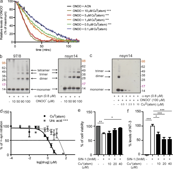
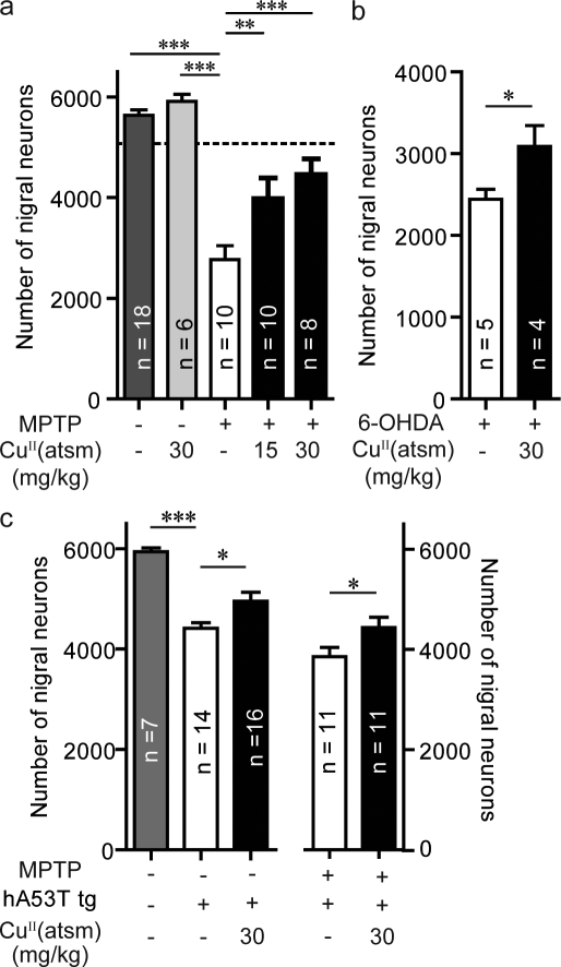
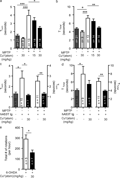
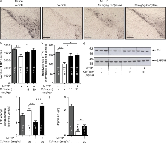
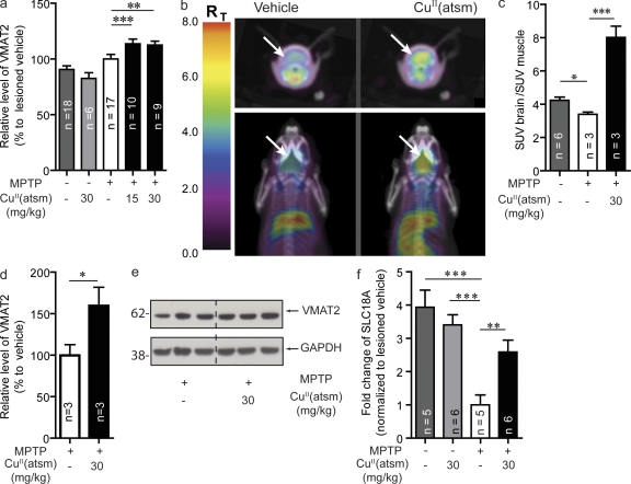
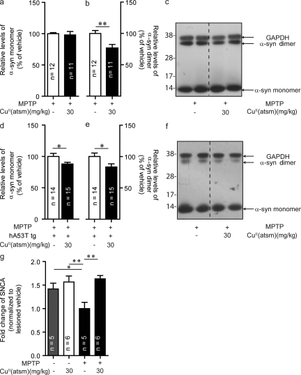
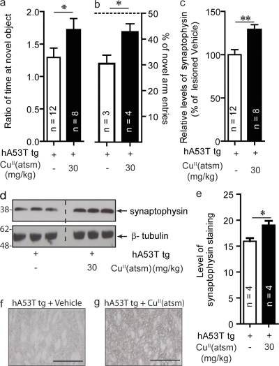
Parkinson's disease (PD) is a progressive, chronic disease characterized by dyskinesia, rigidity, instability, and tremors. The disease is defined by the presence of Lewy bodies, which primarily consist of aggregated α-synuclein protein, and is accompanied by the loss of monoaminergic neurons. Current therapeutic strategies only give symptomatic relief of motor impairment and do not address the underlying neurodegeneration. Hence, we have identified Cu(II)(atsm) as a potential therapeutic for PD. Drug administration to four different animal models of PD resulted in improved motor and cognition function, rescued nigral cell loss, and improved dopamine metabolism. In vitro, this compound is able to inhibit the effects of peroxynitrite-driven toxicity, including the formation of nitrated α-synuclein oligomers. Our results show that Cu(II)(atsm) is effective in reversing parkinsonian defects in animal models and has the potential to be a successful treatment of PD.


Figures

Figure 1.
Figure 1.
CuII(atsm) inhibits ONOO-induced α-synuclein and cell toxicity. (a) Effect of CuII(atsm) on the breakdown of ONOO (500 µM, 302 nm absorbance; CAN, acetonitrile). (b) Effect of increasing concentrations of ONOO on the formation of nitrated α-synuclein oligomers as detected by Western blot using Ab 97/8 (B, left), which detects total α-synuclein, and nsyn14 (B, right), an antibody that specifically detects nitrated α-synuclein. (c) Western blotting with the nsyn14 antibody demonstrates the effect of CuII(atsm) on ONOO-induced nitration of α-synuclein. (d) ELISA using the nsyn14 antibody showing the effect of CuII(atsm) (IC50 of 1 µM) on inhibiting ONOO-induced nitration of α-synuclein compared with uric acid (IC50 of 63 µM), an endogenous ONOO scavenger. (e) MTS cell viability assays on differentiated SH-SY5Y cells after treatment with CuII(atsm) and SIN-1–induced cell toxicity. (f) 3-NT levels in the cell media after CuII(atsm) treatment (as detected by ELISA). For a and d, data are representative of three separate experiments and expressed as mean ± SEM; for statistical analysis, two-way ANOVA was performed with a Bonferroni post-hoc analysis. For e and f, data are representative of three separate experiments and expressed as means ± SEM; for statistical analysis, one-way ANOVA was performed with a Dunnett post-hoc analysis comparing all groups to SIN-1–exposed untreated cells. *, P < 0.05; **, P < 0.01; ***, P < 0.001.
Figure 2.
Figure 2.
CuII(atsm) is neuroprotective in multiple animal models of PD. (a) C57BL/6 mice were lesioned with MPTP (40 mg/kg i.p.). The SNpc count (dotted line) was determined 24 h after MPTP lesioning (when drug treatment commenced). Effect of treatment with CuII(atsm) at both 15 and 30 mg/kg on the survival of SNpc dopaminergic neurons 20 d after lesioning. (b) C57BL/6 mice were lesioned unilaterally with 6-OHDA (intranigral dose). Effect of treatment with 30 mg/kg of CuII(atsm) on the survival of SNpc dopaminergic neurons. (c) Effect of CuII(atsm) (30 mg/kg) treatment of mice overexpressing human A53T (hA53T) α-synuclein, either nonlesioned or lesioned with MPTP on the survival of SNpc dopaminergic neurons. All data are expressed as mean ± SEM; for statistical analysis, one-way ANOVA was performed with a Dunnett post-doc analysis for multisample testing using untreated MPTP-lesioned mice (a), hA53T tg untreated or MPTP-lesioned untreated hA53T tg mice (c), and Student’s t test for two-sample testing (b) as controls. *, P < 0.05; **, P < 0.01; ***, P < 0.001.
Figure 3.
Figure 3.
CuII(atsm) rescues motor impairment in multiple animal models of PD. (a and b) A pole test, which assesses sequential coordinated movement, was performed on MPTP wild-type mice after CuII(atsm) treatment. The ability of an animal to undertake this task is determined by the time the animal takes to turn their body 180 degrees (Tturn; a) and total time to descend the pole (Ttotal; B). (c and d) CuII(atsm) treatment effects on the Tturn in both unlesioned and MPTP-lesioned hA53T α-synuclein tg mice. Differences in Ttotal times were only significant for MPTP-lesioned tg, but not in the nonlesioned tg mice. (e) Effects of CuII(atsm) on the amount of amphetamine-induced rotations in 6-OHDA–lesioned mice. All data are expressed as mean ± SEM; for statistical analysis, one-way ANOVA was performed with a Dunnett post-doc analysis for multisample testing using untreated MPTP-lesioned mice (a and b), untreated hA53T tg mice (c and d), and Student’s t test for two-sample testing (c and e) as controls. *, P < 0.05; **, P < 0.01; ***, P < 0.001.
Figure 4.
Figure 4.
CuII(atsm) treatment increases TH and dopamine. (a) Sections of SNpc stained for TH from unlesioned and MPTP-lesioned mice treated with vehicle, 15 mg/kg CuII(atsm), or 30 mg/kg CuII(atsm). Results were confirmed by stereological counting (b) and Western blotting (c and d). (e) TH results were also confirmed by qPCR. (f) Dopamine levels in CuII(atsm)-treated lesioned animals compared with control lesioned animals. Dopamine levels are expressed relative to tissue weight. All data are expressed as mean ± SEM; for statistical analysis, one-way ANOVA was performed with a Dunnett post-hoc analysis for multisample testing using MPTP-lesioned untreated mice as controls. *, P < 0.05; **, P < 0.01; ***, P < 0001. Bars, 250 µm.
Figure 5.
Figure 5.
CuII(atsm) increases VMAT2 levels in MPTP-lesioned WT mice. (a) VMAT2 levels in young WT (4-mo-old) mice lesioned with MPTP after CuII(atsm) treatment at both 15 and 30 mg/kg doses. (b and c) VMAT2 levels were determined using the specific VMAT2 radioligand 18F-AV133 and microPET imaging in older mice lesioned with MPTP (SUV, standardized uptake value). (d and e) These results were confirmed by Western blot analysis in the striatum. (f) VMAT2 gene (SLC18A) expression after lesioning and CuII(atsm) treatment. All data are expressed as mean ± SEM; for statistical analysis, one-way ANOVA was performed with a Dunnett post-hoc analysis for multi-sample testing using as controls MPTP-lesioned untreated mice (a, c, and f) and Student’s t test for two-sample testing (d) as controls. *, P < 0.05; **, P < 0.01; ***, P < 0.001.
Figure 6.
Figure 6.
CuII(atsm) decreases levels of α-synuclein in the SNpc. (a–c) Effect of CuII(atsm) treatment on α-synuclein levels in MPTP-lesioned WT mice. (d–f) α-Synuclein monomer and dimer levels after treatment of hA53T α-synuclein tg mice lesioned with MPTP. (g) α-Synuclein gene (SNCA) at the transcriptional level in MPTP-lesioned WT mice and after CuII(atsm) treatment. All data are expressed as mean ± SEM; for statistical analysis, one-way ANOVA was performed with a Dunnett post-doc analysis for multisample testing (g) and Student’s t test for 2 sample testing (a, b, d, and e). *, P < 0.05; **, P < 0.01.
Figure 7.
Figure 7.
CuII(atsm) rescues cognitive function in hA53T α-synuclein tg mice. Mice overexpressing human A53T α-synuclein were treated with 30 mg/kg of CuII(atsm) for 20 d beginning at the age of 7 mo. (a) Cognitive performance as tested in a NOR test and in a Y-maze test (b). (c and d) Synaptophysin levels in the striatum as measured by Western blotting. (e–g) Immunoreactivity of synaptophysin in the striatum of hA53T tg mice treated with either vehicle or CuII(atsm). Bars, 250 µm. All data are expressed as mean ± SEM; for statistical analysis, Student’s t test for 2 sample testing was used. *, P < 0.05; **, P < 0.01.

Similar articles

Cited by

References

    1. Alonso A., Rodríguez L.A., Logroscino G., Hernán M.A. 2007. Gout and risk of Parkinson disease: a prospective study. Neurology. 69:1696–1700 10.1212/01.wnl.0000279518.10072.df - DOI - PubMed
    1. Andreadou E., Nikolaou C., Gournaras F., Rentzos M., Boufidou F., Tsoutsou A., Zournas C., Zissimopoulos V., Vassilopoulos D. 2009. Serum uric acid levels in patients with Parkinson’s disease: their relationship to treatment and disease duration. Clin. Neurol. Neurosurg. 111:724–728 10.1016/j.clineuro.2009.06.012 - DOI - PubMed
    1. Baba M., Nakajo S., Tu P.H., Tomita T., Nakaya K., Lee V.M., Trojanowski J.Q., Iwatsubo T. 1998. Aggregation of α-synuclein in Lewy bodies of sporadic Parkinson’s disease and dementia with Lewy bodies. Am. J. Pathol. 152:879–884 - PMC - PubMed
    1. Beckman J.S. 2009. Understanding peroxynitrite biochemistry and its potential for treating human diseases. Arch. Biochem. Biophys. 484:114–116 10.1016/j.abb.2009.03.013 - DOI - PMC - PubMed
    1. Beckman J.S., Beckman T.W., Chen J., Marshall P.A., Freeman B.A. 1990. Apparent hydroxyl radical production by peroxynitrite: implications for endothelial injury from nitric oxide and superoxide. Proc. Natl. Acad. Sci. USA. 87:1620–1624 10.1073/pnas.87.4.1620 - DOI - PMC - PubMed

Publication types

MeSH terms