. 2012 Jun;32(11):2044-53.
doi: 10.1128/MCB.05716-11. Epub 2012 Apr 2.

Deep intron elements mediate nested splicing events at consecutive AG dinucleotides to regulate alternative 3' splice site choice in vertebrate 4.1 genes

Affiliations

Deep intron elements mediate nested splicing events at consecutive AG dinucleotides to regulate alternative 3' splice site choice in vertebrate 4.1 genes

Marilyn K Parra et al. Mol Cell Biol. 2012 Jun.

Abstract

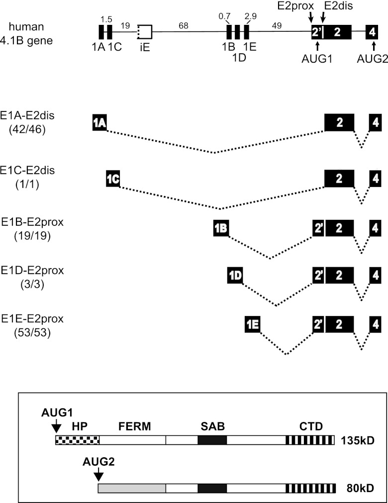
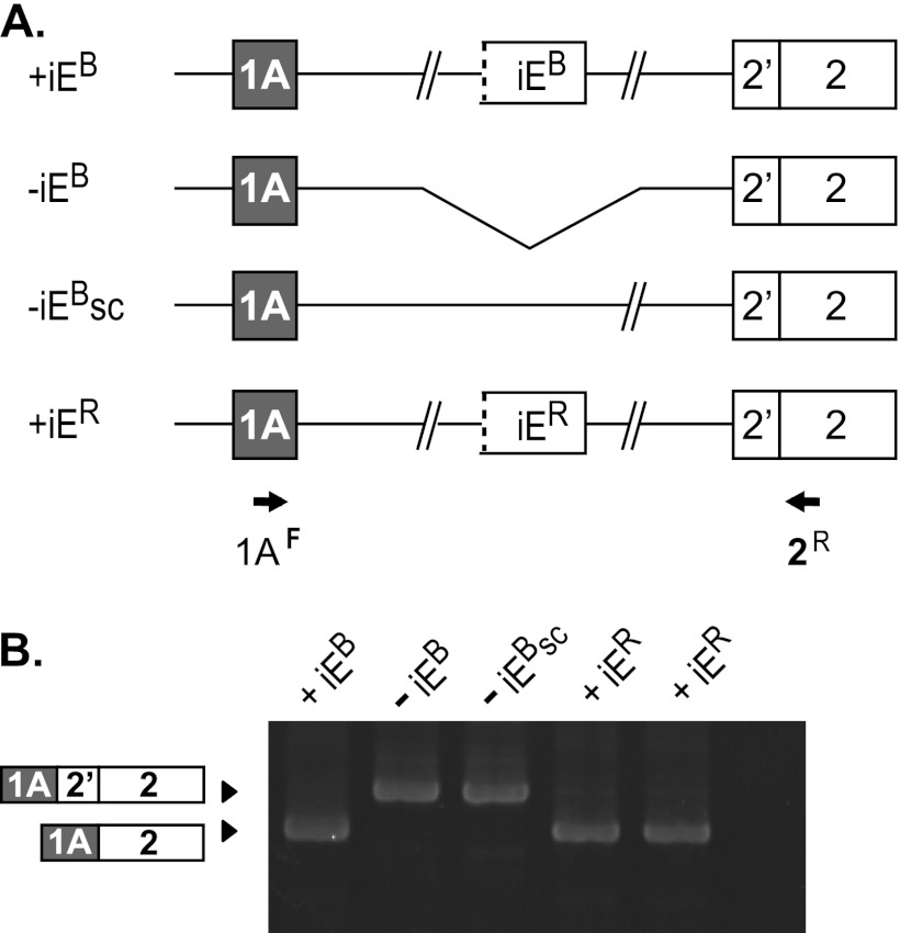
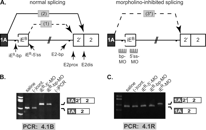
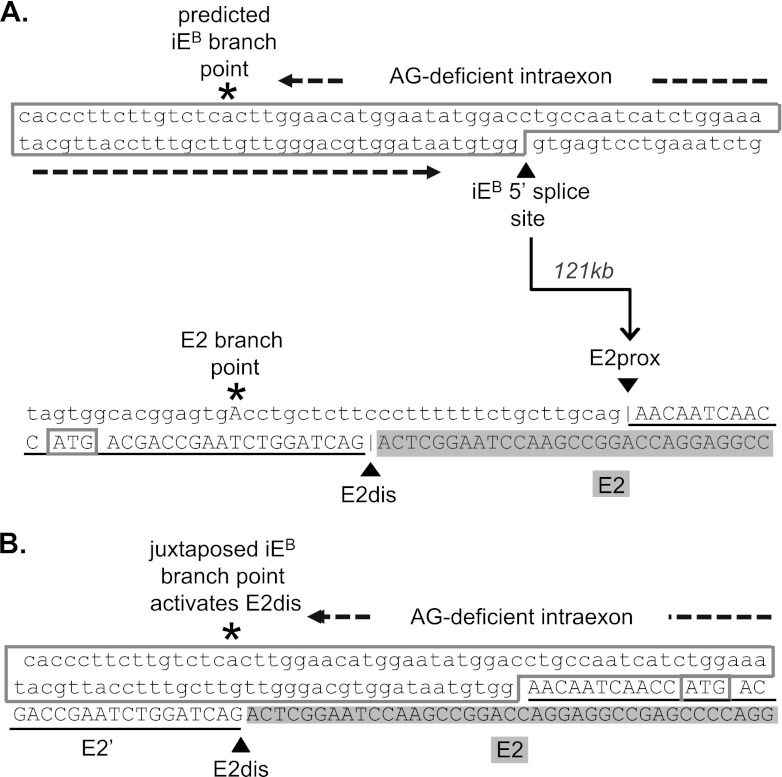
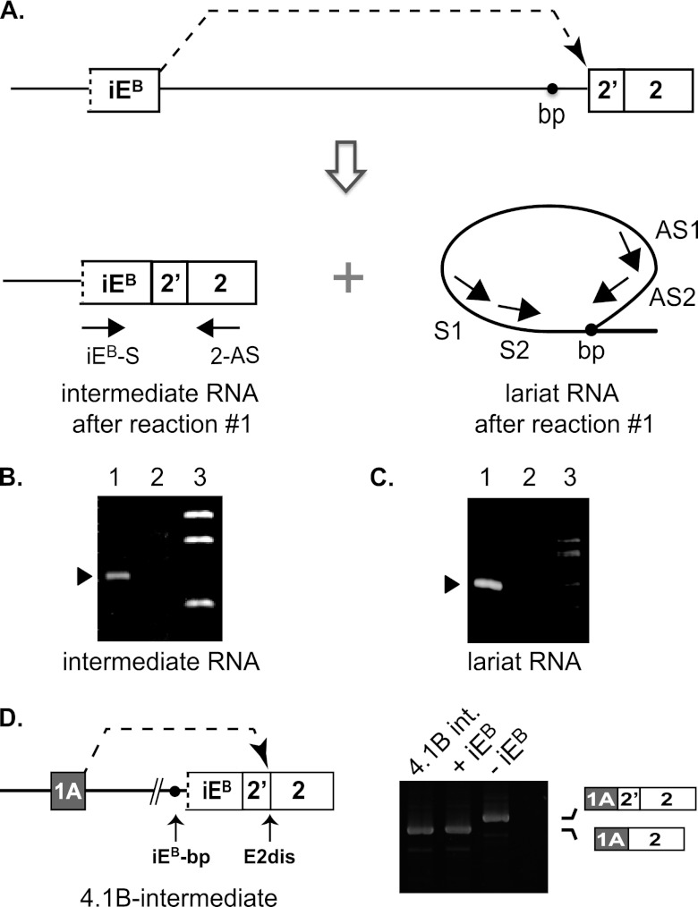
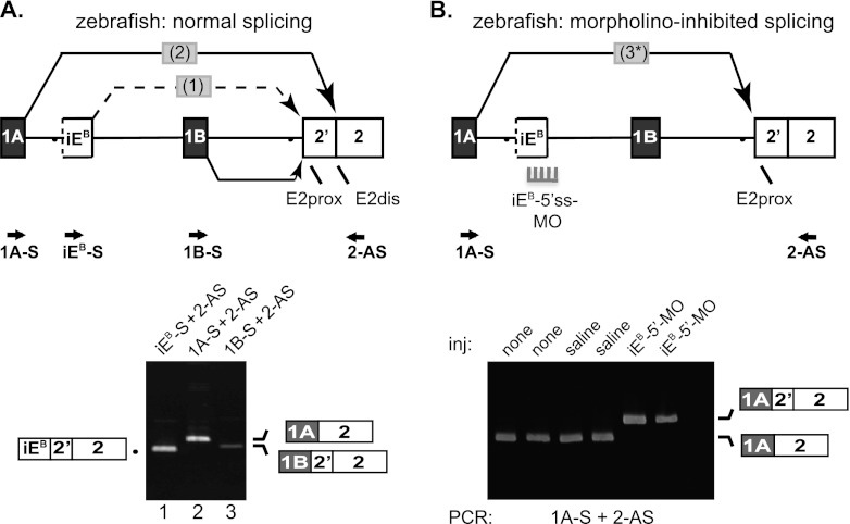
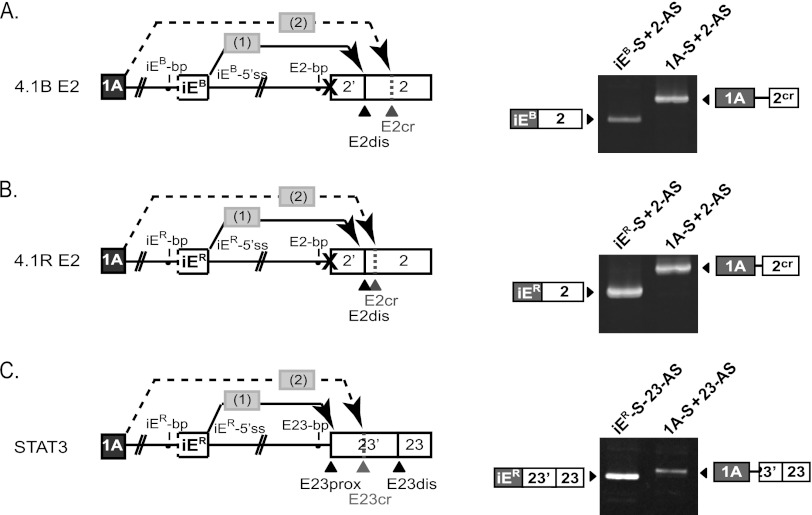
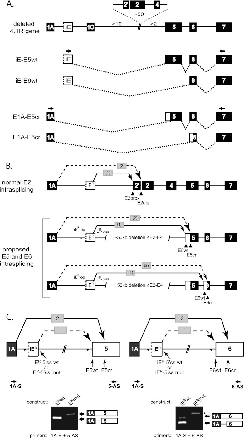
Distal intraexon (iE) regulatory elements in 4.1R pre-mRNA govern 3' splice site choice at exon 2 (E2) via nested splicing events, ultimately modulating expression of N-terminal isoforms of cytoskeletal 4.1R protein. Here we explored intrasplicing in other normal and disease gene contexts and found conservation of intrasplicing through vertebrate evolution. In the paralogous 4.1B gene, we identified ∼120 kb upstream of E2 an ultradistal intraexon, iE(B), that mediates intrasplicing by promoting two intricately coupled splicing events that ensure selection of a weak distal acceptor at E2 (E2dis) by prior excision of the competing proximal acceptor (E2prox). Mutating iE(B) in minigene splicing reporters abrogated intrasplicing, as did blocking endogenous iE(B) function with antisense morpholinos in live mouse and zebrafish animal models. In a human elliptocytosis patient with a mutant 4.1R gene lacking E2 through E4, we showed that aberrant splicing is consistent with iE(R)-mediated intrasplicing at the first available exons downstream of iE(R), namely, alternative E5 and constitutive E6. Finally, analysis of heterologous acceptor contexts revealed a strong preference for nested 3' splice events at consecutive pairs of AG dinucleotides. Distal regulatory elements may control intrasplicing at a subset of alternative 3' splice sites in vertebrate pre-mRNAs to generate proteins with functional diversity.


Figures

Fig 1
Fig 1
Promoter location relative to the iE regulator determines alternative splicing decisions that determine the N-terminal structure of protein 4.1B isoforms. (Top) Exon-intron arrangement at the 5′ end of the 4.1B gene. Numbers indicate intron lengths (in kb) for the human gene. Alternative first exons are indicated E1A-E1E; E2prox and E2dis represent alternative 3′ splice sites in E2; AUG1 and AUG2 represent alternative translation initiation sites. The putative intraexon, iE, is drawn with a broken line on one side to indicate the lack of a functional 3′ splice site. Depicted below the gene model are pre-mRNA splice patterns derived from databases showing that alternative first exons 1A and E1C splice to E2dis, while E1B, 1D, and 1E splice to E2prox. Numbers in parentheses indicate the fraction of all database transcripts initiated at a given promoter that splice to E2 as shown. (Bottom) Larger protein isoforms encoded by transcripts that splice to E2prox and include AUG1, and smaller proteins encoded by transcripts that splice to E2dis so as to skip AUG1 and initiate translation downstream at AUG2. Domains of 4.1R protein: HP, headpiece at N terminus; FERM, 4.1/ezrin/radixin/moesin homology domain; SAB, spectrin-actin binding domain; CTD, C-terminal domain.
Fig 2
Fig 2
The 4.1B intraexon is a powerful splicing regulator required for E1A splicing to E2dis. (A) Splicing reporter constructs either lacking (−iEB) or containing inserts of 0.5 kb encompassing the 4.1B intraexon element (+iEB), additional intron sequence downstream of E1A as a size control (−iEsc), or the 4.1R intraexon (+iER). (B) Splicing analysis of the reporters after transfection into HEK293 cells, using the indicated PCR primers. Structure of the PCR products was confirmed by sequence analysis. Lanes are labeled with names of the constructs.
Fig 3
Fig 3
Splice-blocking vivo-morpholinos validate intraexon regulatory function in full-length endogenous 4.1B pre-mRNA in vivo. (A) (Top) The two nested splicing events (1 and 2) proposed for joining E1A to E2dis (left). Splice-blocking morpholinos targeting the iEB 5′ splice site (5′ss-MO) and branch point (bp-MO) were predicted to abolish iEB function, leading to aberrant one-step splicing (3*) of E1A to E2prox (right). (B) RT-PCR analysis of endogenous mouse kidney 4.1B transcripts with primers located in E1A and E2, using RNA from animals treated with sterile saline, a negative-control vMO [(-)cont], or iEB-specific vMOs against iEB-5′ss and iEB-bp. The first lane shows size standards, and the last lane is an RT-PCR negative control. (C) RT-PCR analysis using 4.1R-specific primers to show that splicing of endogenous mouse kidney 4.1R transcripts was not altered by vMOs against the paralogous 4.1B pre-mRNA. Sources of RNA (indicated above each lane) were isolated from animals injected with sterile saline, with negative-control vMO, with 4.1B-specific vMOs against iEB-5′ss or iEB-bp, or with a 4.1R-specific vMO against iER-5′ss. The first lane shows size standards, and the last lane is an RT-PCR negative control. For both panels B and C, correct splicing of E1A to E2dis was observed in mice treated with sterile saline or control vMOs. Gene-specific vMOs induced aberrant splicing of E1A to E2prox only in the cognate 4.1R or 4.1B pre-mRNA; no cross-regulation of the paralogous pre-mRNA was observed.
Fig 4
Fig 4
Sequences of the 4.1B intraexon, the alternative 3′ splice sites in exon 2, and the composite 3′ splice site formed by juxtaposition of iEB-bp and E2′. (A) The top sequence shows the predicted iEB-branch point and 5′ splice site, with an AG-deficient sequence of 85 nt separating these motifs. The bottom sequence represents the E2 branch point and the two alternative 3′ splice sites at the AG dinucleotides labeled E2prox and E2dis. The translation start site ATG1 is boxed. The arrow indicates the first splicing reaction in which the iEB-5′ss splices, across 121 kb of intron, to acceptor site E2dis. (B) Sequence of the composite 3′ splice site region predicted in the intermediate RNA after splicing of iEB to E2prox.
Fig 5
Fig 5
Analysis of intermediate products generated in the first intrasplicing reaction in 4.1B pre-mRNA. (A) Model showing the expected products of the first intrasplicing event: iEB spliced to E2prox, and a lariat intron released in this reaction. Amplification of the branch point region of the lariat RNA was performed via nested PCRs involving antisense primers (AS1 and AS2) located downstream of the iEB 5′ splice site that loops back to the branch point and two sense primers (S1 and S2) located upstream of the branch point. (B) Gel analysis of the predicted 4.1B intermediate RNA in mouse kidney by RT-PCR analysis using primers in iE and E2. Lane 1, detection of intermediate RNA from normal kidney; lane 2, absence of intermediate RNA from kidney exposed to vMO against the 4.1B iEB-5′ss; lane 3, molecular size standards. (C) Gel analysis of the predicted lariat RNA branch point formed in the first intrasplicing reaction, using the nested primers diagrammed in panel A to detect small amounts of this RNA. Lane 1, product obtained after both nested PCRs; lane 2, no product observed after the first PCR with primers AS1 + S1; lane 3, molecular size standards. (D) (Left) Diagram of a reporter construct used to test whether the predicted splicing intermediate is a bona fide precursor of the fully processed mature E1A-E2dis mRNA. (Right) Analysis of spliced products generated in HEK293 cells transfected with the following reporters: intermediate RNA (4.1B-int), +iEB reporter, shown in Fig. 2, and −iEB reporter, also from Fig. 2. The last lane is a negative RT-PCR control.
Fig 6
Fig 6
Analysis of intrasplicing in the zebrafish 4.1B gene. (A) (Top) 5′ structure of the zebrafish 4.1B gene, including the putative intraexon and two nested splicing reactions predicted to be essential for E1A to E2dis splicing. Locations of PCR primers are indicated. Bottom: RT-PCR analysis of normal zebrafish 4.1B transcripts. Lane 1, intermediate RNA product obtained using primers iEB-S and 2-AS; lane 2, E1A-E2dis splicing revealed using primers 1A-S and 2-AS; lane 3, E1B-E2prox splicing demonstrated using primers 1BF and 2R. B. Top: Model for switch in splice acceptor site usage induced in vivo by inhibition of intraexon function. Splice-blocking MO directed against the intraexon 5′ splice site (5′ss-MO) inhibited splicing events 1 and 2, leading to direct splicing of E1A to E2prox in a single aberrant splicing event 3. Bottom: RT-PCR analysis of duplicate experiments using primers 1A-S and 2-AS to amplify zebrafish RNA prepared from uninjected embryos (none); embryos injected with 0.2 M KCl buffer alone (saline); or injected with 2 or 10 ng of 5′ss-MO against the 4.1B intraexon (iEB-5′-MO).
Fig 7
Fig 7
Intrasplicing splice site selection in heterologous sequence contexts. (A) Mutation of the E2prox AG dinucleotide shifts nested splicing events 1 and 2 from E2prox and E2dis to E2dis and E2cr. The gel shows results of PCR analysis of the nested splicing events using the indicated primers. (B) Mutation of E2prox induces an AG shift in the nested splicing reactions, leading to activation of a cryptic acceptor in E2. As in panel A, the diagram and RT-PCR analysis show the intermediate product from step 1 and final spliced product from step 2. (C) Nested splicing at E23 of the STAT3 gene shows that step 2 of intrasplicing selects the first downstream AG at E23cr rather than the authentic distal acceptor site E23dis.
Fig 8
Fig 8
Misdirected intrasplicing activates cryptic splice sites in a patient with deletion of 4.1R exons 2 to 4. (A) Structure of a reported 4.1R gene deletion and major aberrant splicing events observed at E5 and E6 (4), except that exon 1B in reference is designated here as iER due to its unique properties mediating intrasplicing. Whereas iER in the patient spliced to the normal wild type 3′ splice sites (E5wt and E6wt), E1A spliced exclusively to aberrant cryptic splice sites (E5cr and E6cr). (B) Proposed intrasplicing model to explain aberrant splicing at E5 and E6. Splicing data are consistent with nested splicing events precisely analogous to those reported originally at E2 (top) (25), except that the deletion of E2-E4 shifts iER-mediated intrasplicing to alternative exon E5 and constitutive exon E6. (C) Intrasplicing analysis in minigene splicing reporters containing a functional (iER-5′ss) or inactivated (iER-5′ss mut) intraexon (top) upstream of either E5 or E6. RT-PCR analysis revealed that E1A splices to the normal acceptors at E5 and E6 in the absence of functional iE (lanes iERwt). In contrast, iER-mediated intrasplicing resulted smaller products representing aberrant splicing at cryptic sites, exactly as observed in the patient (lanes iERmut).
Fig 9
Fig 9
The second step of intrasplicing preferentially activates the next downstream AG dinucleotide. The primary sequence of 3′ splice site choice in the first and second steps of intrasplicing for several exons is shown. The second step exhibits a strong preference for activating the next available AG based on position rather than splice site strength.

References

    1. Auboeuf D, Batsche E, Dutertre M, Muchardt C, O'Malley BW. 2007. Coregulators: transducing signal from transcription to alternative splicing. Trends Endocrinol. Metab. 18:122–129 - PubMed
    1. Baek D, Davis C, Ewing B, Gordon D, Green P. 2007. Characterization and predictive discovery of evolutionarily conserved mammalian alternative promoters. Genome Res. 17:145–155 - PMC - PubMed
    1. Baklouti F, et al. 1997. Organization of the human protein 4.1 genomic locus: new insights into the tissue-specific alternative splicing of the pre-mRNA. Genomics 39:289–302 - PubMed
    1. Baklouti F, et al. 2011. Homozygous deletion of EPB41 genuine AUG-containing exons results in mRNA splicing defects, NMD activation and protein 4.1R complete deficiency in hereditary elliptocytosis. Blood Cells Mol. Dis. 47:158–165 - PubMed
    1. Burnette JM, Miyamoto-Sato E, Schaub MA, Conklin J, Lopez AJ. 2005. Subdivision of large introns in Drosophila by recursive splicing at nonexonic elements. Genetics 170:661–674 - PMC - PubMed

Publication types

LinkOut - more resources