. 2012 May 1;188(9):4602-10.
doi: 10.4049/jimmunol.1200021. Epub 2012 Apr 2.

NIK prevents the development of hypereosinophilic syndrome-like disease in mice independent of IKKα activation

Affiliations

NIK prevents the development of hypereosinophilic syndrome-like disease in mice independent of IKKα activation

Hans Häcker et al. J Immunol. .

Abstract

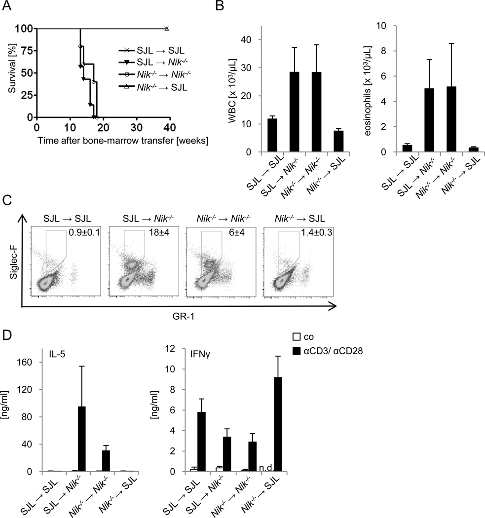
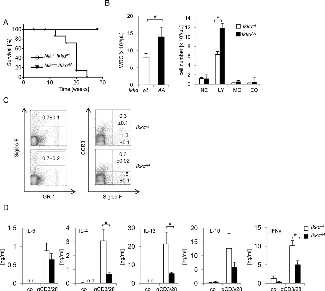
Immune cell-mediated tissue injury is a common feature of different inflammatory diseases, yet the pathogenetic mechanisms and cell types involved vary significantly. Hypereosinophilic syndrome (HES) represents a group of inflammatory diseases that is characterized by increased numbers of pathogenic eosinophilic granulocytes in the peripheral blood and diverse organs. On the basis of clinical and laboratory findings, various forms of HES have been defined, yet the molecular mechanism and potential signaling pathways that drive eosinophil expansion remain largely unknown. In this study, we show that mice deficient of the serine/threonine-specific protein kinase NF-κB-inducing kinase (NIK) develop a HES-like disease, reflected by progressive blood and tissue eosinophilia, tissue injury, and premature death at around 25-30 wk of age. Similar to the lymphocytic form of HES, CD4(+) T cells from NIK-deficient mice express increased levels of Th2-associated cytokines, and eosinophilia and survival of NIK-deficient mice could be prevented completely by genetic ablation of CD4(+) T cells. Experiments based on bone marrow chimeric mice, however, demonstrated that inflammation in NIK-deficient mice depended on radiation-resistant tissues, implicating that NIK-deficient immune cells mediate inflammation in a nonautonomous manner. Surprisingly, disease development was independent of NIK's known function as an IκB kinase α (IKKα) kinase, because mice carrying a mutation in the activation loop of IKKα, which is phosphorylated by NIK, did not develop inflammatory disease. Our data show that NIK activity in nonhematopoietic cells controls Th2 cell development and prevents eosinophil-driven inflammatory disease, most likely using a signaling pathway that operates independent of the known NIK substrate IKKα.


Figures

FIGURE 1
FIGURE 1. NIK deficient mice develop a progressive inflammatory disease characterized by blood and tissue eosinophilia and premature death
A, Thickening of the eyelids, fur loss and skin inflammation as typically observed in Nik−/− mice at around 12 weeks of age. B, Survival curve of Nik+/+ and Nik−/− mice. C, Age at disease onset as determined by thickening of eyelids, fur loss or skin inflammation in male and female mice. D, White blood cell count (WBC) and differential cell count of peripheral blood of 12–16 weeks old Nik+/+ and Nik−/− mice. NE, neutrophilic granulocytes; LY, lymphocytes; MO, monocytes; EO, eosinophils; n=9, data represent mean±SEM. E, Flow cytometry analysis of spleens of 12 week old Nik+/+ and Nik−/− mice for immature (GR-1int Siglec-F+ CCR3) and mature (GR-1int Siglec-F+ CCR3+) eosinophils. n=5, data represent mean±SEM. F, Representative H&E stain and eosinophil-specific anti-MBP stain of indicated organs of 16 week old Nik+/+ and Nik−/− mice. All images are shown at 40x magnification. G, Representative T-cell specific anti-CD3 stain of the lung and liver of 16 week old Nik+/+ and Nik−/− mice shown at 40x magnification.
FIGURE 2
FIGURE 2. Th2-biased T-cell phenotype and reduced CD4+/ CD8+ T-cell ratio in NIK-deficient mice
A, Purified CD4+ T-cells from 6–16 week old Nik+/+ and Nik−/− mice were left untreated (co) or stimulated with αCD3/αCD28 for 72 hours and cytokine levels were determined by ELISA. n=9, data represent mean±SEM, *p≤0.005, **p≤0.001, ***p≤0.0005. B, Purified CD4+ T-cells from 2 week old Nik+/+ and Nik−/− mice were stimulated with αCD3/αCD28 in triplicate wells and analyzed as described in A. C, Flow cytometry analysis of splenocytes from 6–12 week old Nik+/+ and Nik−/− mice. Total splenocytes (left panel), CD3+-gated cells (middle panel) or CD3+ CD4+-gated cells (right panel) are shown. n=3, data represent mean±SEM. CD8+ NIK+/+ vs. CD8+ NIK−/− *** p=0.0005, CD4+ NIK+/+ vs. CD4+ NIK−/− *** p= 0.0002.
FIGURE 3
FIGURE 3. Inflammation in Nik−/− mice depends on CD4+ T-cells
A, Nik−/− mice were crossed to Rag1−/− mice and survival of offspring was monitored for 40 weeks. Nik−/− Rag+/+ (n=5), Nik−/− Rag−−/− (n=10). B, Histological analysis based on H&E and MBP staining of lung and liver of 16 week old Nik−/− Rag+/+ and Nik−/− Rag−/− mice. Representative data obtained from 6 mice are shown. C, Nik−/− mice were crossed to MhcII−/− mice and survival of offspring was monitored for 40 weeks. Nik−/− MhcII+/+ (n=10), Nik−/−MhcII−/− (n=10). D, Histological analysis based on H&E and MBP staining of lung and liver of 16 week old Nik−/− MhcII+/+ and Nik−/−MhcII−/− mice. Representative data obtained from 3 mice are shown.
FIGURE 4
FIGURE 4. Radiation-resistant cells in NIK-deficient mice trigger inflammatory disease
A, Lethally irradiated wt (SJL) and Nik−/− mice were reconstituted with bone marrow cells of the indicated genotypes, and survival was monitored for 40 weeks. n=4–7. B, White blood cell count and differential cell count of peripheral blood of bone marrow chimeric mice described in A at 8 weeks after bone marrow transfer. n=4–7, data represent mean±SEM. C, Splenocytes of bone marrow chimeric mice described in A were analyzed by flow cytometry 8 weeks after transfer. n=2–3, chimerism was >90%. Live cells were gated and are represented as mean±SEM. D, Purified CD4+ T-cells from bone marrow chimeric mice described in A were isolated 8 weeks after transfer and either left untreated (co) or stimulated with αCD3/αCD28 for 72 hours, and IL-5 and IFNγ levels were determined by ELISA. n=2–3, data represent mean±SEM.
FIGURE 5
FIGURE 5. Disease in Nik−/− mice proceeds independent of IKKα phosphorylation
A, Nik−/− mice were crossed to IkkαAA/AA (IkkαAA) mice. Survival of Nik−/− Ikkαwt/wt (n=7), Nik−/− IkkαAA/AA (n=9) mice was monitored over 30 weeks. B, White blood cell count (WBC) and differential cell count of peripheral blood of 6–16 week old Ikkαwt/wt (wt) and IkkαAA/AA (AA) mice. NE, neutrophilic granulocytes; LY, lymphocytes; MO, monocytes; EO, eosinophils; n=7, data represent mean±SEM. C, Flow cytometry analysis of splenocytes from 16 week old Ikkαwt/wt and IkkαAA/AA mice for immature (GR-1int Siglec-F+ CCR3) and mature (GR-1int Siglec-F+ CCR3+) eosinophils, n=3, data represent mean±SEM. D, Purified CD4+ T-cells from 12–16 week old IKKαwt/wt and IKKαAA/AA mice were left untreated (co) or stimulated with αCD3/αCD28 for 72 hours and cytokine levels were determined by ELISA. n=5, data represent mean±SEM, *p≤0.005.

References

    1. Simon HU, Rothenberg ME, Bochner BS, Weller PF, Wardlaw AJ, Wechsler ME, Rosenwasser LJ, Roufosse F, Gleich GJ, Klion AD. Refining the definition of hypereosinophilic syndrome. J. Allergy Clin. Immunol. 126:45–49. - PMC - PubMed
    1. Gotlib J. World Health Organization-defined eosinophilic disorders: 2011 update on diagnosis, risk stratification, and management. Am. J. Hematol. 86:677–688. - PubMed
    1. Ogbogu PU, Bochner BS, Butterfield JH, Gleich GJ, Huss-Marp J, Kahn JE, Leiferman KM, Nutman TB, Pfab F, Ring J, Rothenberg ME, Roufosse F, Sajous MH, Sheikh J, Simon D, Simon HU, Stein ML, Wardlaw A, Weller PF, Klion AD. Hypereosinophilic syndrome: a multicenter, retrospective analysis of clinical characteristics and response to therapy. J. Allergy Clin. Immunol. 2009;124:1319–1325. e1313. - PMC - PubMed
    1. Gleich GJ, Leiferman KM. The hypereosinophilic syndromes: current concepts and treatments. Br. J. Haematol. 2009;145:271–285. - PubMed
    1. Roufosse F, Cogan E, Goldman M. Recent advances in pathogenesis and management of hypereosinophilic syndromes. Allergy. 2004;59:673–689. - PubMed

Publication types

MeSH terms