. 2012 May 1;188(9):4506-15.
doi: 10.4049/jimmunol.1200202. Epub 2012 Apr 2.

The Asp299Gly polymorphism alters TLR4 signaling by interfering with recruitment of MyD88 and TRIF

Affiliations

The Asp299Gly polymorphism alters TLR4 signaling by interfering with recruitment of MyD88 and TRIF

Leandra Figueroa et al. J Immunol. .

Abstract

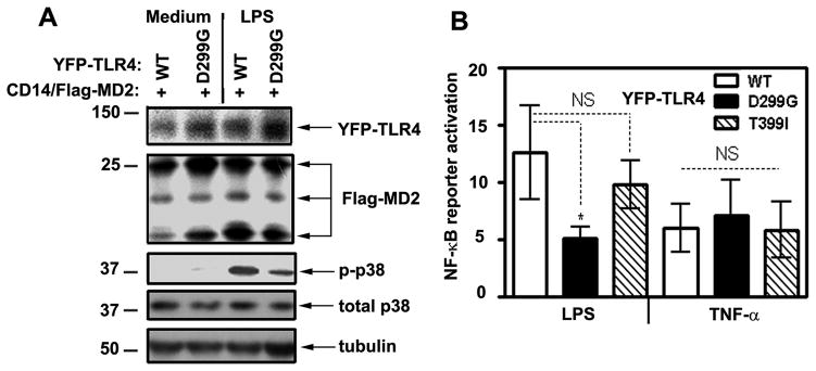
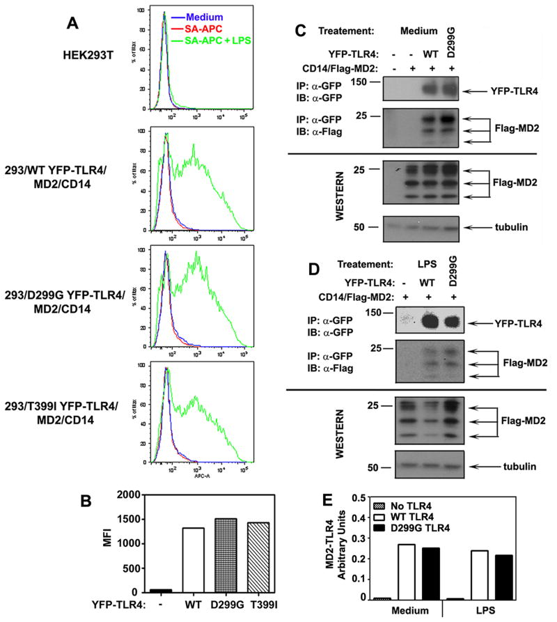
Asp(299)Gly (D299G) and, to a lesser extent, Thr(399)Ile (T399I) TLR4 polymorphisms have been associated with gram-negative sepsis and other infectious diseases, but the mechanisms by which they affect TLR4 signaling are unclear. In this study, we determined the impact of the D299G and T399I polymorphisms on TLR4 expression, interactions with myeloid differentiation factor 2 (MD2), LPS binding, and LPS-mediated activation of the MyD88- and Toll/IL-1R resistance domain-containing adapter inducing IFN-β (TRIF) signaling pathways. Complementation of human embryonic kidney 293/CD14/MD2 transfectants with wild-type (WT) or mutant yellow fluorescent protein-tagged TLR4 variants revealed comparable total TLR4 expression, TLR4-MD2 interactions, and LPS binding. FACS analyses with anti-TLR4 Ab showed only minimal changes in the cell-surface levels of the D299G TLR4. Cells transfected with D299G TLR4 exhibited impaired LPS-induced phosphorylation of p38 and TANK-binding kinase 1, activation of NF-κB and IFN regulatory factor 3, and induction of IL-8 and IFN-β mRNA, whereas T399I TLR4 did not cause statistically significant inhibition. In contrast to WT TLR4, expression of the D299G mutants in TLR4(-/-) mouse macrophages failed to elicit LPS-mediated induction of TNF-α and IFN-β mRNA. Coimmunoprecipitation revealed diminished LPS-driven interaction of MyD88 and TRIF with the D299G TLR4 species, in contrast to robust adapter recruitment exhibited by WT TLR4. Thus, the D299G polymorphism compromises recruitment of MyD88 and TRIF to TLR4 without affecting TLR4 expression, TLR4-MD2 interaction, or LPS binding, suggesting that it interferes with TLR4 dimerization and assembly of intracellular docking platforms for adapter recruitment.


Figures

Figure 1
Figure 1. The presence of the D299G polymorphism impairs the ability of TLR4 to mediate LPS-induced p38 phosphorylation and NF-κB activation
A, HEK293 cells stably expressing WT or D299G YFP-TLR4 were co-transfected with pcDNA3-CD14 and pEFBOS-Flag-MD2. Cells were treated for 30 min with medium or 100 ng/ml LPS, and cell lysates were analyzed by immunoblotting with Abs against GFP (YFP-TLR4), Flag (Flag-MD2), p-p38, total p38, and tubulin. The data of a representative (n=3) experiment are shown. B, HEK293T cells transiently transfected with WT, D299G or T399I YFP-TLR4 species, along with pcDNA3-CD14 and pEFBOS-Flag-MD2 were co-transfected with pELAM-Luc and pTK-Renilla-Luc. Cells were treated for 5 h with medium, 100 ng/ml LPS or TNF-α, and cell lysates were analyzed for firefly and Renilla Luc activities. The summary of five experiments (mean ± SD) is depicted. *p<0.05; NS: non-significant.
Figure 2
Figure 2. The impact of the D299G and T399I polymorphisms on total and cell surface expression of TLR4
HEK293 cells stably transfected with expression vectors encoding WT, D299G or T399I YFP-TLR4 were co-transfected with pcDNA3-CD14 and pEFBOS-Flag-MD2. Cells transfected with pcDNA3, designated as “(−)”, were used as a negative control. After recovery, total TLR4 expression was determined by confocal microscopy (A) and FACS analysis (B, top panel), using the YFP fluorescence intensity (FITC channel) as a readout. TLR4 cell surface expression was determined by staining of cells with HTA125-PE (anti-TLR4 Ab) or isotype control IgG-PE, followed by FACS analyses of PE fluorescence (B, bottom panel). C: Whole cell lysates from the respective cell transfectants were subjected to Western blot analyses, using antibodies against GFP, Flag, and tubulin. The results of a representative (n=3) experiment are shown.
Figure 3
Figure 3. LPS binding to the TLR4/CD14/MD2 complex and TLR4-MD2 interactions in HEK293/CD14/MD2 cells expressing WT, D299G or T399I YFP-TLR4 species
A, HEK293 cells stably expressing WT, D299G or T399I YFP-TLR4 species were transiently transfected with pcDNA3-CD14 and pEFBOS-Flag-MD2. After recovery for 20 h, cells were incubated for 30 min at 37° C with medium or biotinylated LPS (1μg/ml), washed, stained with SA-APC (1μg/ml), and analyzed by FACS to detect LPS binding (APC fluorescence). B: Quantification of FACS results (mean fluorescence intensity, MFI) shown in (A), using the FlowJo program. HEK293T cells were transiently transfected with vectors encoding WT or D299G YFP-TLR4 species, along with pcDNA3-CD14/pEFBOS-Flag-MD2. After recovery, cells were treated with medium (C) or stimulated with 100 ng/ml LPS (D), cell lysates were immunoprecipitated with anti-GFP antibody (YFP-TLR4 pull-down), and analyzed by immunoblotting with anti-Flag Ab (to determine Flag-MD2 interactions with YFP-TLR4s). Tubulin immunoblot was used to control protein loading. The data of a representative experiment (n=3) are shown. E: Densotometric quantification of the data shown in (C, D). The results are presented as arbitrary units representing ratios of normalized densitometric values of MD2-TLR4 complexes detected in samples transfected with pcDNA3 (background), or vectors expressing WT or D299G YFP-TLR4. Normalization of densitometric values was calculated as a/b/c/d, where a, b, c and d correspond to the densitometric values of MD2-TLR4, total TLR4, total MD2 and tubulin, respectively.
Figure 4
Figure 4. Compromised LPS-mediated recruitment of MyD88 to D299G TLR4
HEK293 cells transiently (A) or stably (B) expressing WT or D299G YFP-TLR4 were co-transfected with pcDNA3-CD14, pEFBOS-HA-MD2, and either pcDNA3-AU1 or pcDNA3. After recovery, cells were treated with medium or 100 ng/ml LPS for 1 min (A) or for the indicated time course (B), cell lysates were prepared, and YFP-TLR4 proteins were immunoprecipitated with anti-GFP Ab or isotype control IgG, followed by immunoblot analyses of the immune complexes with anti-GFP or anti-AU1 Abs. Whole cell lysates were examined by immunoblotting, using the indicated antibodies. C and D: densitometirc quantification of the data shown in A and B. Densities of the bands representing MyD88 associated with TLR4 were normalized by total TLR4 and by total MD2 values and were expressed as arbitrary units. Numbers indicate inhibition (%) of LPS-inducible MyD88-TLR4 values in D299G YFP-TLR4-transfected cells compared to WT YFP-TLR4 transfectants. Shown are the results of a representative (n=3) experiment.
Figure 5
Figure 5. The impact of the D299G and T399I TLR4 polymorphisms on LPS-induced expression of MyD88-dependent cytokine genes
HEK293 transient (A) and stable (B) transfectants expressing WT, D299G or T399I YFP-TLR4 were co-transfected with pcDNA3-CD14/pEFBOS-Flag-MD2. C, C57BL/6 (TLR4+/+) and TLR4−/− primary BMDMs were nucleofected with plasmids encoding eGFP (a negative control), WT or D299G YFP-TLR4, as indicated. After recovery for 24 h. cells were treated for 3 h with medium, LPS or TNF-α (100 ng/ml each), RNA was isolated, reverse-transcribed, and analyzed by real-time qPCR with primers specific for human HPRT and IL-8 (A, B); mouse HPRT and TNF-α (C), and human TLR4 (D). The results of a representative experiment (n = 3) are depicted. *p<0.05; ns: not statistically significant.
Figure 6
Figure 6. The D299G polymorphism impairs the ability of TLR4 to recruit TRIF in response to LPS stimulation
293/TLR4 stable cell lines expressing WT or D299G YFP-TLR4 species were co-transfected with pcDNA3-CD14, pEFBOS-HA-MD2, and pEFBOS-Flag-TRIF. After recovery for 20 h, cells were treated with medium or stimulated for the indicated times with 100 ng/ml LPS, cell lysates were prepared and immunoprecipitated with anti-GFP or anti-Flag Abs. Immune complexes were analyzed by immunoblotting with anti-GFP, anti-Flag, or isotype control IgG Abs to determine total TLR4 expression and amounts of TRIF recruited to TLR4 proteins. The data of a representative (n=3) experiments are shown.
Figure 7
Figure 7. LPS-mediated phosphorylaiton of TBK-1 and IRF3, activation of p125-Luc and induction of IFN-β mRNA in cells expressing WT or D299G YFP-TLR4
A, HEK293T cells were transiently transfected with plasmids encoding WT or D299G YFP-TLR4, along with pcDNA3-CD14 and pEFBOS-Flag-MD2. After recovery, cells were treated with 100 ng/ml LPS for 30 min and lysates were analyzed by immunoblotting with anti-GFP (YFP-TLR4), anti-Flag (Flag-MD2), anti-p-TBK-1, anti-total TBK-1, and anti-tubulin Abs. B, HEK293 cells stably expressing YFP-TLR4 WT or D299G were co-transfected with pcDNA3-CD14 and pEFBOS-HA-MD2. After recovery, cells were treated for with 100 ng/ml LPS for 15 min, and cell lysates were prepared and analyzed by immunoblotting with Abs against GFP (YFP-TLR4), p-IRF3 and total IRF3, and anti-HA-HRP Ab (HA-MD2). C, HEK293T cells were transiently transfected with WT, D299G or T399I YFP-TLR4 species, along with pcDNA3-CD14 and pEFBOS-Flag-MD2. In addition, cells were co-transfected with p125-Luc and pTK-Renilla-Luc. D, HEK293 cells stably expressing WT or D299G YFP-TLR4 species were co-transfected with pcDNA3-CD14 and pEFBOS-Flag-MD2. After recovery, cells were stimulated for 5 h with LPS or TNF-α (100 ng/ml each). Cell lysates were analyzed for firefly and Renilla luciferase activities. E, HEK293T cells were transiently transfected with WT, D299G or T399I YFP-TLR4 species, along with pcDNA3-CD14 and pEFBOS-Flag-MD2. F, WT (TLR4+/+) and TLR4−/− iBMDMs were transfected as shown. After recovery, cells were stimulated for 3 h with 100 ng/ml LPS. RNA was isolated, reverse-transcribed and examined by real-time qPCR with primers specific for human HPRT and IFN-β (E) or mouse HPRT and IFN-β (F). Shown are results (mean ± SD) of the summary of three independent experiments (C and D) and a representative experiment (n=3, A, B, E and F). *p<0.05;**p<0.01; ns: not significant.

Similar articles

Cited by

References

    1. Medzhitov R, Preston-Hurlburt P, Janeway CA., Jr A human homologue of the Drosophila Toll protein signals activation of adaptive immunity. Nature. 1997;388:394–397. - PubMed
    1. Beutler B. Microbe sensing, positive feedback loops, and the pathogenesis of inflammatory diseases. Immunol Rev. 2009;227:248–263. - PMC - PubMed
    1. Schattgen SA, Fitzgerald KA. The PYHIN protein family as mediators of host defenses. Immunol Rev. 2011;243:109–118. - PubMed
    1. Davis BK, Wen H, Ting JP. The inflammasome NLRs in immunity, inflammation, and associated diseases. Annu Rev Immunol. 2011;29:707–735. - PMC - PubMed
    1. Onoguchi K, Yoneyama M, Fujita T. Retinoic acid-inducible gene-I-like receptors. J Interferon Cytokine Res. 2011;31:27–31. - PubMed

Publication types

MeSH terms