. 2012 May;26(5):819-32.
doi: 10.1210/me.2011-1140. Epub 2012 Apr 3.

Bile acids down-regulate caveolin-1 in esophageal epithelial cells through sterol responsive element-binding protein

Affiliations

Bile acids down-regulate caveolin-1 in esophageal epithelial cells through sterol responsive element-binding protein

Elke Prade et al. Mol Endocrinol. 2012 May.

Abstract

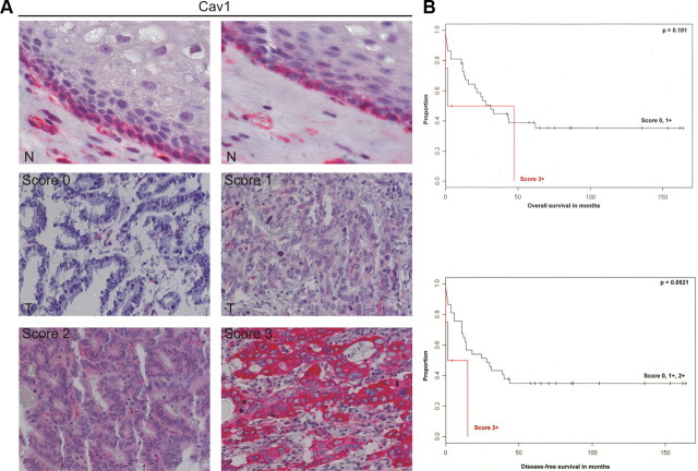
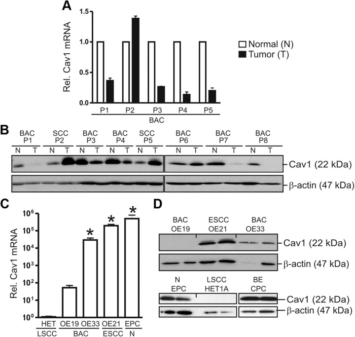
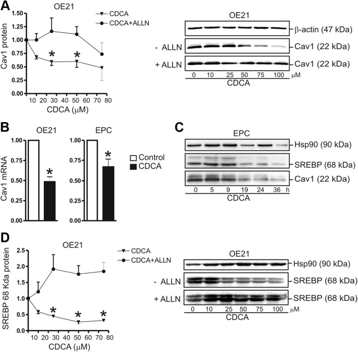
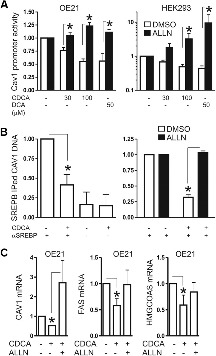
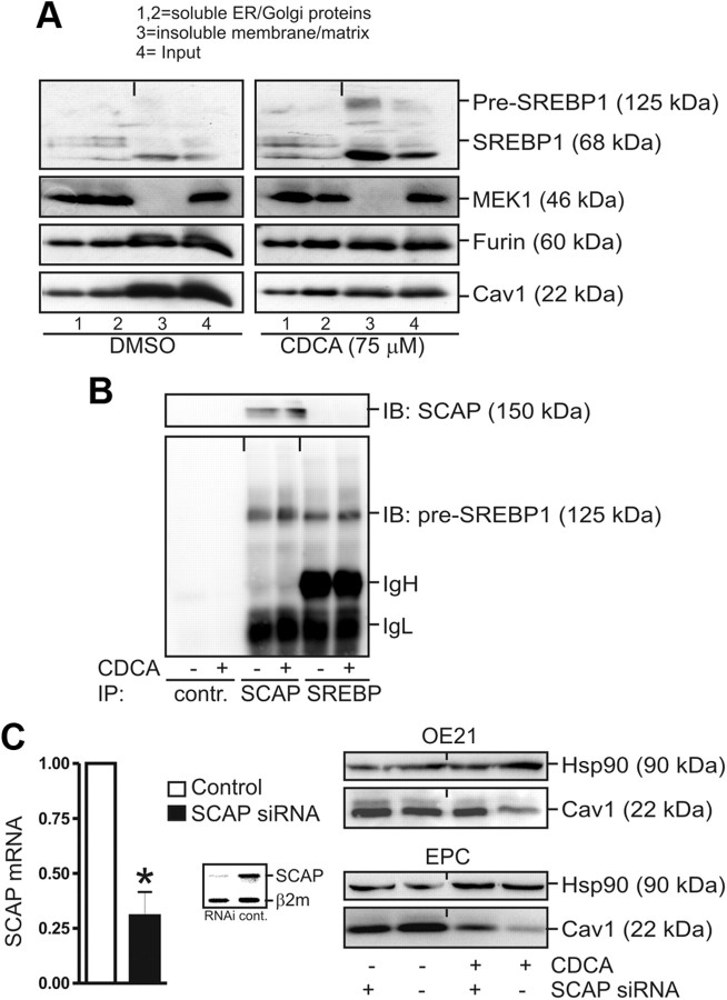
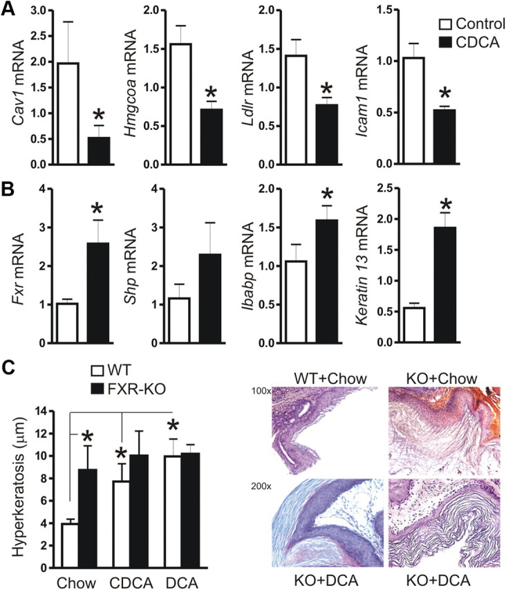
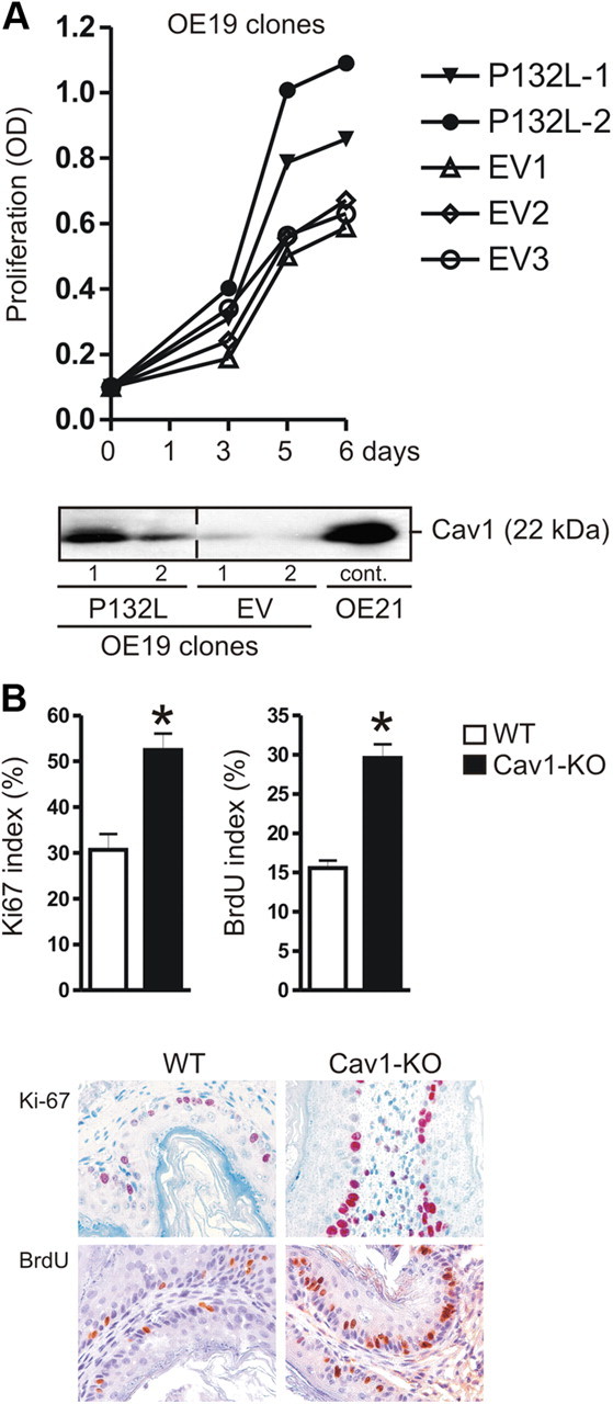
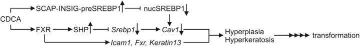
Bile acids are synthesized from cholesterol and are major risk factors for Barrett adenocarcinoma (BAC) of the esophagus. Caveolin-1 (Cav1), a scaffold protein of membrane caveolae, is transcriptionally regulated by cholesterol via sterol-responsive element-binding protein-1 (SREBP1). Cav1 protects squamous epithelia by controlling cell growth and stabilizing cell junctions and matrix adhesion. Cav1 is frequently down-regulated in human cancers; however, the molecular mechanisms that lead to this event are unknown. We show that the basal layer of the nonneoplastic human esophageal squamous epithelium expressed Cav1 mainly at intercellular junctions. In contrast, Cav1 was lost in 95% of tissue specimens from BAC patients (n = 100). A strong cytoplasmic expression of Cav1 correlated with poor survival in a small subgroup (n = 5) of BAC patients, and stable expression of an oncogenic Cav1 variant (Cav1-P132L) in the human BAC cell line OE19 promoted proliferation. Cav1 was also detectable in immortalized human squamous epithelial, Barrett esophagus (CPC), and squamous cell carcinoma cells (OE21), but was low in BAC cell lines (OE19, OE33). Mechanistically, bile acids down-regulated Cav1 expression by inhibition of the proteolytic cleavage of 125-kDa pre-SREBP1 from the endoplasmic reticulum/Golgi apparatus and nuclear translocation of active 68-kDa SREBP1. This block in SREBP1's posttranslational processing impaired transcriptional activation of SREBP1 response elements in the proximal human Cav1 promoter. Cav1 was also down-regulated in esophagi from C57BL/6 mice on a diet enriched with 1% (wt/wt) chenodeoxycholic acid. Mice deficient for Cav1 or the nuclear bile acid receptor farnesoid X receptor showed hyperplasia and hyperkeratosis of the basal cell layer of esophageal epithelia, respectively. These data indicate that bile acid-mediated down-regulation of Cav1 marks early changes in the squamous epithelium, which may contribute to onset of Barrett esophagus metaplasia and progression to BAC.


Figures

Fig. 1.
Fig. 1.
Expression of Cav1 in human BAC patients. A, Cav1 is lost in the majority of BAC patients. IHC detection of Cav1 protein in human nonneoplastic esophageal tissue and BAC specimens (n = 100) using a monoclonal antibody; N, normal squamous epithelium (top panels) with Cav1 at cell junctions; T, BAC (lower panels). The staining scores for Cav1 in BAC tumor cells were correlated to disease-free and overall survival. Score: 0, negative; +1, positive (weak); +2, positive (moderate); +3, positive (strong). B, Strong cytoplasmic expression of Cav1 (score 3+) in a small subgroup of BAC patients (n = 5) is a negative prognostic factor for survival. Kaplan-Meier curves are presented for overall and disease-free survival.
Fig. 2.
Fig. 2.
Expression of Cav1 in human EC cell lines. A and B, Expression of Cav1 mRNA (A) and protein (B) in human esophageal cancer (EC) detected by RT-qPCR and WB. Cav1 is reduced in BAC as compared with matched normal tissue and SCC tissues. CT values from RT-qPCR were normalized to β2-microglobulin (β2M) and presented as -fold ± se (n = 5 patients); *, P < 0.05; N (Normal) vs. T (Tumor). Total tissue lysates were normalized to protein content and compared with β-actin. Representative WB gels are shown. SCC, squamous cell carcinoma; BAC, Barrett adenocarcinoma. C and D, Detection of Cav1 mRNA (C) and protein (D) in human EC cell lines. CT values from RT-qPCRs were normalized to β2-microglobulin (β2M) and presented as -fold ± se (n = 3); *, P < 0.05 vs. OE19. Total cell lysates were normalized to protein content and compared with β-actin. Representative WB gels are shown. OE19 and OE33, BAC cell lines; OE21, SCC cell line; EPC, immortalized esophageal squamous epithelial cell line; CPC, Barrett esophagus (metaplasia) cell line; HET1A, control squamous epithelial cell line. Rel, Relative.
Fig. 3.
Fig. 3.
Bile acids down-regulate Cav1 and SREBP1 in normal human esophageal and SCC cells. A, Concentration-dependent reduction of Cav1 protein by CDCA. OE21 cells were treated for 30 h, in serum-free medium, with the concentration indicated of CDCA, in the presence and absence of the protease inhibitor 10 μm ALLN. O.D. values from bands in WB gels are expressed as -fold ± se (n = 3); *, P < 0.05 CDCA vs. DMSO. Quantitative analyses (left) are presented with representative WB (right). B, Down-regulation of Cav1 mRNA by CDCA. OE21 and EPC cells were treated as in panel A. CT values from RT-qPCRs were normalized to β2-microglobulin (β2M) and presented as -fold ± se (n = 3); *, P < 0.05 CDCA vs. DMSO. C, Time-dependent reduction of Cav1 and 68-kDa active SREBP1 proteins by CDCA. EPC cells were treated for the indicated times with 75 μm CDCA. Representative WB are shown. D, Concentration-dependent decrease of 68-kDa active SREBP1 protein. OE21 cells were incubated for 30 h with the indicated concentrations of CDCA, in the presence and absence of 10 μm ALLN, respectively. Quantitative analyses (left) and representative WB (right) are shown. Hsp, Heat shock protein.
Fig. 4.
Fig. 4.
Bile acids inhibit activation of the human Cav1 promoter by active 68-kDa SREBP1. A, CDCA inhibits transactivation of the human Cav1 promoter. OE21 and HEK293 (control) cells were transiently transfected with a Cav1 promoter-driven luciferase reporter plasmid and treated for 30 h with CDCA (30 μm and 100 μm) and 50 μm DCA, with and without 10 μm ALLN, respectively. Luciferase activity was normalized to protein content and presented as -fold ± se (n = 3); *, P < 0.05 CDCA or DCA vs. DMSO. B, CDCA blocks binding of SREBP1 to sterol-response elements (SRE) in the human Cav1 promoter. Cells were treated as in panel A. IP was performed on cell lysates with rabbit polyclonal SREBP1 antiserum or control IgG. The CT values from genomic qPCR of IP-ed DNA were normalized to the CT values of input DNA and are expressed as -fold ± se. (n = 3) change of pulldown by CDCA compared with control; *, P < 0.05 CDCA vs. DMSO. C, Down-regulation of SREBP1 target genes by CDCA is reversed by ALLN, an inhibitor of active 68-kDa SREBP1's catabolism. OE21 cells were incubated for 30 h with 100 μm CDCA in the absence or presence of 10 μm ALLN. CT values from RT-qPCR were normalized to β2-microglobulin (β2M) and presented as -fold ± se (n = 3); *, P < 0.05 CDCA vs. DMSO or ALLN. FAS, Fatty acid synthase.
Fig. 5.
Fig. 5.
Bile acids inhibit nuclear accumulation of active 68-kDa SREBP1 involving SCAP. A, Sucrose gradient ultracentrifugation. OE21 cells were grown to confluency in 15-cm dishes and treated for 30 h with 100 μm CDCA or DMSO, respectively. Fractions were subjected to WB: 1,2, soluble ER and Golgi membrane proteins; 3, insoluble membrane, cytoskeleton, and matrix proteins; 4, input control. Note the reduced Cav1 protein and enhanced membrane retention (lane 3) of 125-kDa pre-SREBP1 and 68-kDa SREBP1 upon CDCA treatment. B, CDCA promotes interaction of pre-SREBP1 with SCAP. OE21 cells were grown and treated as in panel A, and CoIP was performed using SCAP and SREBP1 polyclonal antibodies. Representative WB are shown. C, SCAP is required for CDCA-mediated down-regulation of Cav1. OE21 cells were transiently transfected with siRNA oligonucleotides against SCAP or control siRNA, respectively, and treated with 75 μm CDCA for 30 h. RT-qPCR analyses (left) and representative WB (right) are shown. CT values from RT-qPCR were normalized to β2-microglobulin (β2M) and presented as -fold ± se (n = 3); *, P < 0.05 SCAP-siRNA vs. control-siRNA. Insert, Agarose gel from RT-PCR showing SCAP mRNA knockdown by siRNA. hsp90, Heat shock protein 90; IB, immunoblotting; IP, immunoprecipitation; MEK, MAPK kinase.
Fig. 6.
Fig. 6.
Bile acids down-regulate Cav1 also in vivo and activate FXR. A, Down-regulation of mRNAs encoding cell adhesion proteins (Cav1, Icam1) and SREBP1-target genes (Hmgcoas, Ldlr) in C57BL/6 mice on a 7-d chow diet enriched with 1% (wt/wt) CDCA as compared with littermates on control chow. CT values from RT-qPCR on total RNA extracted from resected esophagi were normalized to β2-microglobulin (β2M) and presented as means ± se (n = 5 per group); *. P < 0.05 CDCA vs. chow. B, Up-regulation of mRNA for FXR-target genes (Fxr, Shp, Ibabp, Keratin13). Data are presented as in panel A. C, FXR-KO mice show hyperkeratosis of the squamous epithelium. C57BL/6 WT and FXR-KO mice (n = 5 per group) were fed a chow diet with or without 1% (wt/wt) CDCA or 0.2% (wt/wt) DCA for 8 wk, respectively. H&E stainings from paraffin sections of esophageal and forestomach squamous epithelial tissue was evaluated for the thickness of the keratinizing sloughing tissue. The diameter of the keratin layer was measured (five random fields per mouse) and presented as μm ± se (n = 5 per group); *, P < 0.05 WT vs. FXR-KO; chow vs. CDCA/DCA. Quantitative analyses (left) are shown together with representative histology (right); magnifications ×100 and ×200.
Fig. 7.
Fig. 7.
Cav1-deficiency increases proliferation in vitro and in vivo. A, The oncogenic dominant-negative Cav1 variant P132L promotes proliferation of OE19 cells. A (bottom panel), Cav1 protein expression in stably transfected OE19 cells. Representative WB are shown. A (top panel), Time course of proliferation of OE19 clones. OD values from colorimetric MTT (3-(4,5-dimethylthiazol-2-yl)-2,5-diphenyltetrazolium bromide) assays are presented as means ± se (n = 3) of Cav1-transfected (P132L) cells compared with EV-transfected cells; *, P < 0.05 P132L vs. EV. B, Cav1-KO mice display hyperplasia of the squamous epithelium. Quantitative analyses of IHC for Ki-67 and BrdU uptake; magnification, ×200. The ratio of positive to total nuclei in the basal layer of the squamous epithelium of forestomach and esophageal tissue was calculated as % ± se (n = 5 per genotype); *, P < 0.05 WT vs. Cav1-KO (both at an age of 4 months).
Fig. 8.
Fig. 8.
Signaling model of bile acid-induced changes in esophageal squamous epithelia. CDCA or other bile acids (such as DCA in the refluxate) bind to the nuclear bile acid receptor FXR and activate expression of its cognate target gene SHP. SHP itself represses Srebp1c gene transcription. In parallel, CDCA mimics high-sterol conditions and inhibits proteolytic cleavage and nuclear accumulation of active 68-kDa SREBP1 at target gene promoters. Defective SREBP1 transactivation leads to reduced expression of target genes including Cav1. Other FXR-target genes such as Icam-1 and Keratin 13 may contribute to the observed hyperkeratosis phenotype of FXR-KO mice. Loss of Cav1 leads to squamous cell hyperplasia in Cav1-KO mice and may activate EGFR signaling and destabilize cell-cell and cell-matrix contacts. In sum, the SCAP/SREBP1 and the FXR/SHP pathways are expected to converge in vivo, to evoke early molecular changes in the esophageal squamous epithelium, which may ultimately facilitate BE metaplasia and transformation, preneoplastic lesions in the progression to BAC.

References

    1. Lagergren J. 2005. Adenocarcinoma of oesophagus: what exactly is the size of the problem and who is at risk? Gut 54 Suppl 1:i1–5 - PMC - PubMed
    1. Shaheen NJ. 2005. Advances in Barrett's esophagus and esophageal adenocarcinoma. Gastroenterology 128:1554–1566 - PubMed
    1. Anderson LA , Watson RG , Murphy SJ , Johnston BT , Comber H , Mc Guigan J , Reynolds JV , Murray LJ. 2007. Risk factors for Barrett's oesophagus and oesophageal adenocarcinoma: results from the FINBAR study. World J Gastroenterol 13:1585–1594 - PMC - PubMed
    1. Flook NW , Wiklund I. 2007. Accounting for the effect of GERD symptoms on patients' health-related quality of life: supporting optimal disease management by primary care physicians. Int J Clin Pract 61:2071–2078 - PMC - PubMed
    1. Perez MJ , Briz O. 2009. Bile-acid-induced cell injury and protection. World J Gastroenterol 15:1677–1689 - PMC - PubMed

Publication types

MeSH terms