. 2012 Apr 1;26(7):693-704.
doi: 10.1101/gad.182758.111.

Quantitative functions of Argonaute proteins in mammalian development

Affiliations

Quantitative functions of Argonaute proteins in mammalian development

Dongmei Wang et al. Genes Dev. .

Abstract

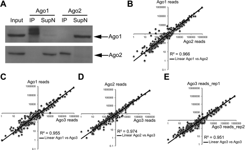
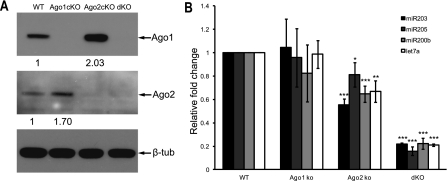
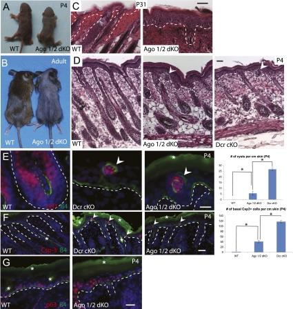
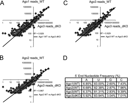
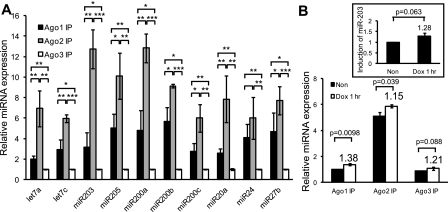
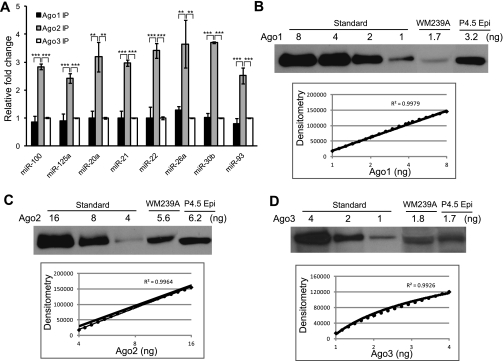
Argonaute proteins (Ago1-4) are essential components of the microRNA-induced silencing complex and play important roles in both microRNA biogenesis and function. Although Ago2 is the only one with the slicer activity, it is not clear whether the slicer activity is a universally critical determinant for Ago2's function in mammals. Furthermore, functional specificities associated with different Argonautes remain elusive. Here we report that microRNAs are randomly sorted to individual Argonautes in mammals, independent of the slicer activity. When both Ago1 and Ago2, but not either Ago1 or Ago2 alone, are ablated in the skin, the global expression of microRNAs is significantly compromised and it causes severe defects in skin morphogenesis. Surprisingly, Ago3 is able to load microRNAs efficiently in the absence of Ago1 and Ago2, despite a significant loss of global microRNA expression. Quantitative analyses reveal that Ago2 interacts with a majority of microRNAs (60%) in the skin, compared with Ago1 (30%) and Ago3 (<10%). This distribution is highly correlated with the abundance of each Argonaute, as quantified by shotgun proteomics. The quantitative correlation between Argonautes and their associated microRNAs is conserved in human cells. Finally, we measure the absolute expression of Argonaute proteins and determine that their copy number is ~1.4 × 10(5) to 1.7 × 10(5) molecules per cell. Together, our results reveal a quantitative picture for microRNA activity in mammals.


Figures

Figure 1.
Figure 1.
Argonautes associate with a similar pool of miRNAs. (A) Specification and efficiency characterization of Ago1 and Ago2 antibodies. Ago1 or Ago2 antibody specifically pulls down Ago1 or Ago2 in IP, depleting the proteins in the supernatant. (B) Comparison of miRNA cloning frequency of Ago1 and Ago2 IP. (C) Comparison of miRNA cloning frequency of Ago1 and Ago3 IP. (D) Comparison of miRNA cloning frequency of Ago2 and Ago3 IP. (E) Two biological duplicates of Ago3 IP show high correlations, confirming the reproducibility of small RNA cloning.
Figure 2.
Figure 2.
Genetic deletion of individual Argonautes results in a quantitative loss of global miRNA expression in the skin. (A) Validation of individual or combinatorial Ago conditional knockout (cKO) by Western blotting. (dKO) Ago1/2 double cKO. Ago1 expression increases in Ago2 cKO, whereas Ago2 expression increases in Ago1 cKO. (B) The reduction of miRNA expression in Ago1 cKO, Ago2 cKO, and Ago1/2 dKO. (*) P < 0.05; (**) P < 0.01; (***) P < 0.001; n = 3.
Figure 3.
Figure 3.
Combinatorial deletion of Ago1 and Ago2 significantly impairs the miRNA pathway and results in severe skin defects. (A) At birth, Ago1/2 dKO is slightly smaller than wild-type (WT) littermate. (B) Ago1/2 dKO lives to adulthood. Adult dKO shows sparse hair coat with a body size similar to wild-type littermate. (C) Degenerated hair follicles and thickened epidermis in adult dKO. (D) Abnormal hair follicles and cysts (arrowhead) are detected in dKO, as indicated by H&E staining. (E) Validation of the hair follicle original of the cyst by Lef1 immunofluorescence staining. Ago1/2 dKO have less cyst than Dicer cKO (Dcr cKO). (F) Apoptosis (marked by cleaved caspase-3 staining) is detected in dKO epidermis, comparable with Dcr cKO with a lower frequency. (G) Expansion of p63-positive cells in dKO epidermis. The dotted lines mark the boundary between the epidermis and dermis. White asterisks denote autofluorescence. Bar, 20 μm. (*) P < 0.05; n = 3.
Figure 4.
Figure 4.
miRNAs are randomly distributed among individual Argonautes. (A) Comparison of miRNA cloning frequency of Ago1 IP in wild-type (WT) cells with Ago3 IP in dKO cells shows high correlations. (B) Comparison of miRNA cloning frequency of Ago2 IP in wild-type cells with Ago3 IP in dKO cells shows high correlations. (C) Comparison of miRNA cloning frequency of Ago3 IP in wild-type cells with Ago3 IP in dKO cells shows high correlations. (D) Cloning frequencies show no distinguishable preferences for 5′-nucleotide identity in individual Ago-IPs in wild-type cells compared with Ago3 IP in dKO cells.
Figure 5.
Figure 5.
Quantitative distributions of miRNAs among Argonautes are determined by their expression level in the skin. (A) By IP-qPCR, Ago2 associates with the most miRNAs, followed by Ago1, then Ago3. Shown are 10 abundantly expressed miRNAs in mouse epidermis. When normalized to Ago3-associated miRNAs (set as 1.0 U), Ago1 associates with an average of 3.8 U of miRNAs, and Ago2 associates with an average of 8.5 U of miRNAs. (*) P < 0.05; (**) P < 0.01; (***) P < 0.001; n = 3. (B) Newly synthesized miR-203 distributes proportionally to three Argonautes in cultured keratinocytes. (Inset) The overall miR-203 level is increased to 1.28-fold after 1 h of induction. P-value is shown (n = 3).
Figure 6.
Figure 6.
Quantitative distributions of miRNAs among Argonautes are determined by their expression level in human melanoma cells. (A) Quantitative analysis of miRNAs binding to three Argonautes in human melanoma cell line WM239A. When normalized to Ago3-associated miRNAs (set as 1.0 U), Ago1 associates with an average of 0.95 U of miRNAs, and Ago2 associates with an average of 2.8 U of miRNAs. No significant difference was observed between Ago1 IP and Ago3 IP for any miRNA. (**) P < 0.01; (***) P < 0.001; n = 3. (B) Absolute quantification of Ago1 expression in WM239A and P4.5 epidermis using synthetic Ago1 protein. (C) Absolute quantification of Ago2 expression in WM239A and P4.5 epidermis using synthetic Ago2 protein. (D) Absolute quantification of Ago3 expression in WM239A and P4.5 epidermis using synthetic Ago3 protein. (P4.5 Epi) P4.5 epidermis.
Figure 7.
Figure 7.
Overexpression of individual Ago proteins rescues the growth defects in Ago1/2 dKO cells. (A) Schematic illustration of the experimental design. (B) Most of GFP-infected Ago1/2fl/fl keratinocytes die after the removal of Ago1/2 by AdCre. In contrast, overexpression of either Ago2 or Ago3 rescues the growth defects. (***) P < 0.001, compare with the GFP control; n = 4.

References

    1. Andl T, Murchison EP, Liu F, Zhang Y, Yunta-Gonzalez M, Tobias JW, Andl CD, Seykora JT, Hannon GJ, Millar SE 2006. The miRNA-processing enzyme dicer is essential for the morphogenesis and maintenance of hair follicles. Curr Biol 16: 1041–1049 - PMC - PubMed
    1. Azuma-Mukai A, Oguri H, Mituyama T, Qian ZR, Asai K, Siomi H, Siomi MC 2008. Characterization of endogenous human Argonautes and their miRNA partners in RNA silencing. Proc Natl Acad Sci 105: 7964–7969 - PMC - PubMed
    1. Babiarz JE, Ruby JG, Wang Y, Bartel DP, Blelloch R 2008. Mouse ES cells express endogenous shRNAs, siRNAs, and other Microprocessor-independent, Dicer-dependent small RNAs. Genes Dev 22: 2773–2785 - PMC - PubMed
    1. Bartel DP 2009. MicroRNAs: Target recognition and regulatory functions. Cell 136: 215–233 - PMC - PubMed
    1. Cheloufi S, Dos Santos CO, Chong MM, Hannon GJ 2010. A dicer-independent miRNA biogenesis pathway that requires Ago catalysis. Nature 465: 584–589 - PMC - PubMed

Publication types

LinkOut - more resources