Review
. 2012 May 25;287(22):17905-13.
doi: 10.1074/jbc.R112.356006. Epub 2012 Apr 2.

Hedgehog secretion and signal transduction in vertebrates

Affiliations
Review

Hedgehog secretion and signal transduction in vertebrates

Kaitlyn E Ryan et al. J Biol Chem. .

Abstract

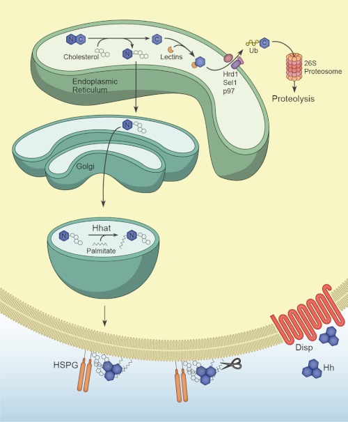
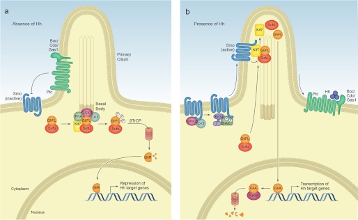
Signaling by the Hedgehog (Hh) family of secreted proteins is essential for proper embryonic patterning and development. Dysregulation of Hh signaling is associated with a variety of human diseases ranging from developmental disorders such as holoprosencephaly to certain forms of cancer, including medulloblastoma and basal cell carcinoma. Genetic studies in flies and mice have shaped our understanding of Hh signaling and revealed that nearly all core components of the pathway are highly conserved. Although many aspects of the Drosophila Hh pathway are conserved in vertebrates, mechanistic differences between the two species have begun to emerge. Perhaps the most striking divergence in vertebrate Hh signaling is its dependence on the primary cilium, a vestigial organelle that is largely absent in flies. This minireview will provide an overview of Hh signaling and present recent insights into vertebrate Hh secretion, receptor binding, and signal transduction.


Figures

FIGURE 1.
FIGURE 1.
Hh processing and release. Hh precursor peptides 45 kDa in size undergo a cholesterol-dependent autocatalytic cleavage in the endoplasmic reticulum to generate a cholesterol-modified N-terminal fragment (Hh-N, denoted by N) and a 25-kDa C-terminal fragment (Hh-C, denoted by C). Hh-C is recognized by the lectins OS-9 and XTP3 and ubiquitylated by the ubiquitin ligase Hrd1 and its partner Sel1. Ubiquitylated Hh-C is moved into the cytosol by the p97 ATPase and subsequently degraded by the proteasome. Cholesterol-modified Hh-N enters the secretory pathway, where the acyltransferase Hhat catalyzes the covalent attachment of palmitate to the N-terminal cysteine. Dually lipidated Hh is targeted to the cell membrane, where cholesterol facilitates the assembly of multimeric Hh complexes possibly by tethering Hh to the membrane and promoting interactions with HS proteoglycans (HSPG). Prior to its release, N- and C-terminal peptides may be cleaved by membrane-proximal proteases such as those belonging to the ADAM family, resulting in the removal of both lipid moieties. The 12-pass transmembrane protein Disp facilitates the release of Hh multimers into the extracellular environment, although the mechanistic details of this process are not well understood. Ub, ubiquitin.
FIGURE 2.
FIGURE 2.
Regions of Shh important for receptor binding and multimerization. Shown is the structure of human SHH-N (non-cholesterol-modified N-terminal fragment; Protein Data Bank code 3M1N (99)). Residues in green (Glu-72, Arg-73, and Lys-75) mediate electrostatic interactions between Hh monomers and are required for multimerization (38). Arg-73 is the vertebrate equivalent of Drosophila Lys-132, the mutation of which results in decreased long-range signaling in the imaginal disc (26). Residues in yellow (His-133, His-134, His-140, His-180, and His-182) are important for Ptc binding (note that His-140 and His-182 coordinate with zinc). Residues in red (Lys-32, Arg-33, Arg-34, Lys-37, and Lys-38) form the Cardin-Weintraub motif and interact with HS. Note how the N terminus extends away from the globular domain of SHH-N; some of these residues may be cleaved in the formation of active Shh multimers (see text).
FIGURE 3.
FIGURE 3.
SHH-N receptor binding involves the Zn2+ coordination site. a, structure of human SHH-N in complex with HIP (Protein Data Bank code 3HO5 (39)). The L2 loop in the β-propeller domain of HIP interacts with SHH-N. b, HIP binds the pseudo-active site in SHH-N, and Asp-383 completes the tetrahedral coordination of Zn2+ in SHH-N. Inset, His-140, His-142, and Arg-147 of SHH-N coordinate Zn2+. Note that the Zn2+ coordination site is also required for binding to PTC, and PTC likely binds SHH in a manner similar to HIP (see text).
FIGURE 4.
FIGURE 4.
Vertebrate Hh signal transduction. a, in the absence of ligand, the 12-pass transmembrane protein Ptc localizes to the primary cilium base and maintains Smo in an inactive conformation. Gli-FL transcription factors complex with Sufu. Sufu sequesters Gli-FL in the cytosol and stabilizes the protein. Sufu and the kinesin-4 family member Kif7 promote the phosphorylation of C-terminal residues in Gli-FL by PKA, GSK3β, and CK1α, which may occur at the basal body of the primary cilium. Phosphorylated Gli-FL is recognized by the E3 ubiquitin ligase βTrCP, resulting in ubiquitylation and proteasomal degradation of C-terminal residues to generate a truncated N-terminal transcriptional repressor (Gli-R) that inhibits Hh target gene transcription. b, in the presence of ligand, Hh binding to Ptc causes Ptc to exit the cilium and relieves its inhibition of Smo. Smo is phosphorylated by CK1α and GRK2, inducing a conformational change and enabling β-arrestin (β-Arr)- and Kif3a-dependent transport into the cilium. Within the cilium, activated Smo promotes the disassembly of Sufu-Gli complexes. Kif7 also localizes to the cilium in the presence of Hh and likely assists Smo in this disassembly. Gli-FL accumulates in the tip of the cilium and is shuttled into the nucleus, perhaps on cytoplasmic microtubules. Within the nucleus, Gli-FL receives additional modifications that convert it to a labile transcriptional activator (Gli-A) that activates Hh target genes. Gli-A is subsequently degraded in a manner that requires the cullin-3-based adaptor Spop.

References

    1. Ingham P. W., McMahon A. P. (2001) Hedgehog signaling in animal development: paradigms and principles. Genes Dev. 15, 3059–3087 - PubMed
    1. Ingham P. W., Nakano Y., Seger C. (2011) Mechanisms and functions of Hedgehog signaling across the metazoa. Nat. Rev. Genet 12, 393–406 - PubMed
    1. Goetz S. C., Anderson K. V. (2010) The primary cilium: a signaling center during vertebrate development. Nat. Rev. Genet. 11, 331–344 - PMC - PubMed
    1. Pedersen L. B., Rosenbaum J. L. (2008) Intraflagellar transport (IFT) role in ciliary assembly, resorption, and signaling. Curr. Top. Dev. Biol. 85, 23–61 - PubMed
    1. Ocbina P. J., Eggenschwiler J. T., Moskowitz I., Anderson K. V. (2011) Complex interactions between genes controlling trafficking in primary cilia. Nat. Genet. 43, 547–553 - PMC - PubMed

Substances

LinkOut - more resources