. 2012 Apr 17;109(16):E916-25.
doi: 10.1073/pnas.1121471109. Epub 2012 Apr 2.

Biotinylation of lysine method identifies acetylated histone H3 lysine 79 in Saccharomyces cerevisiae as a substrate for Sir2

Affiliations

Biotinylation of lysine method identifies acetylated histone H3 lysine 79 in Saccharomyces cerevisiae as a substrate for Sir2

Poonam Bheda et al. Proc Natl Acad Sci U S A. .

Abstract

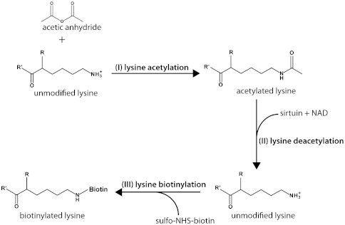
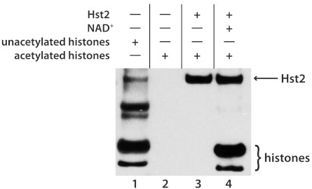
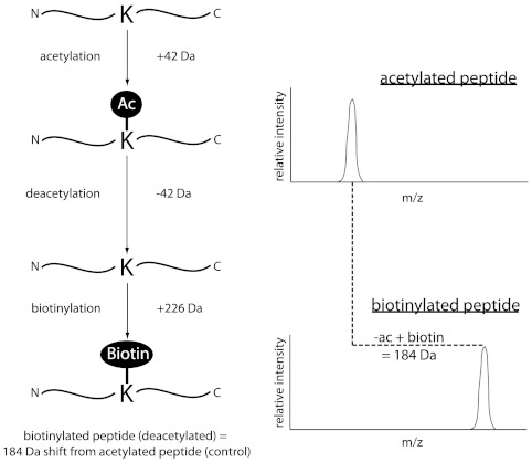
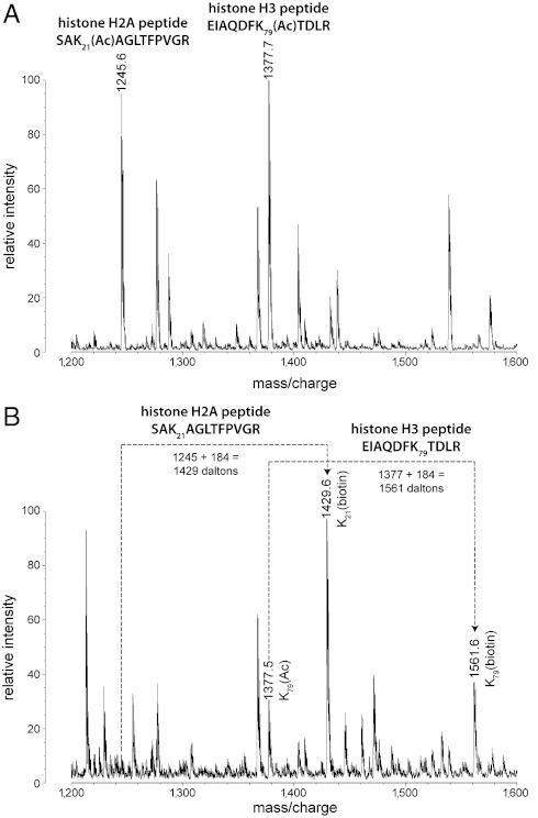
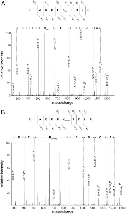
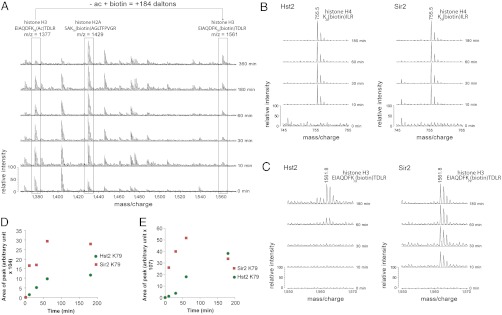
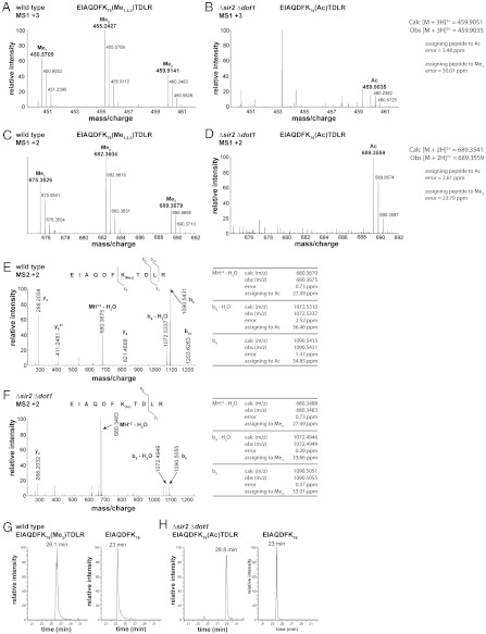
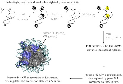
Although the biological roles of many members of the sirtuin family of lysine deacetylases have been well characterized, a broader understanding of their role in biology is limited by the challenges in identifying new substrates. We present here an in vitro method that combines biotinylation and mass spectrometry (MS) to identify substrates deacetylated by sirtuins. The method permits labeling of deacetylated residues with amine-reactive biotin on the ε-nitrogen of lysine. The biotin can be utilized to purify the substrate and identify the deacetylated lysine by MS. The biotinyl-lysine method was used to compare deacetylation of chemically acetylated histones by the yeast sirtuins, Sir2 and Hst2. Intriguingly, Sir2 preferentially deacetylates histone H3 lysine 79 as compared to Hst2. Although acetylation of K79 was not previously reported in Saccharomyces cerevisiae, we demonstrate that a minor population of this residue is indeed acetylated in vivo and show that Sir2, and not Hst2, regulates the acetylation state of H3 lysine 79. The in vitro biotinyl-lysine method combined with chemical acetylation made it possible to identify this previously unknown, low-abundance histone acetyl modification in vivo. This method has further potential to identify novel sirtuin deacetylation substrates in whole cell extracts, enabling large-scale screens for new deacetylase substrates.


Conflict of interest statement

The authors declare no conflict of interest.

Figures

Fig. 1.
Fig. 1.
Overview of the biotinyl-lysine reaction scheme for detection of sirtuin-mediated deacetylation. (I) Lysines and N termini are acetylated with acetic anhydride. (II) Sirtuin-mediated NAD+-dependent deacetylation removes acetyl groups to regenerate unmodified lysines. (III) Deacetylated lysines are biotinylated with sulfo-NHS-biotin.
Fig. 2.
Fig. 2.
Biotinylated proteins detected by blotting with streptavidin-HRP. (Lane 1) Unmodified histones were labeled with biotin at free lysines and N termini. (Lanes 2 and 3) Acetylated histones were not labeled because they did not have any free amines. (Lane 4) Acetylated histones in the presence of sirtuin and NAD+ were deacetylated, and these free lysines were biotinylated, allowing them to be detected by the streptavidin-HRP.
Fig. 3.
Fig. 3.
Schematic depicting changes in molecular weight and mass/charge peak shifts in MALDI-TOF as a result of deacetylation and biotinylation. The molecular weight of a peptide including a lysine is increased by 42 Da by acetylation. Deacetylation removes this 42 Da modification, and biotinylation adds 226 Da. Deacetylated, biotinylated lysines therefore result in a net change of 184 Da compared to acetylated lysines.
Fig. 4.
Fig. 4.
Sample MALDI mass spectrum of Hst2-deacetylated histones. (A) MALDI-TOF spectrum of histone peptide peaks in a control reaction lacking NAD+. (B) MALDI mass spectrum of a deacetylation reaction containing Hst2, NAD+, and histones shows mass shifting of peptides that are deacetylated and biotinylated in comparison to A.
Fig. 5.
Fig. 5.
Sample tandem mass spectra of the doubly charged, histone H3 peptide (aa 73–83), EIAQDFK79TDLR. (A) Mass spectrum of the control sample of histone incubated with only Hst2 shows this peptide with acetylated K79. (B) Mass spectrum of the histone H3 peptide biotinylated at K79. The sample was treated with Hst2 in the presence of NAD+, resulting in the replacement of the acetyl group on K79 with a biotin tag. There is therefore a 92 Da mass increase of the precursor ion and a 184 Da increase in mass between the y4 and y5 ions.
Fig. 6.
Fig. 6.
Histone H3 K79 is preferentially deacetylated by Sir2 in comparison to Hst2. (A) MALDI mass spectra of time courses of Sir2-mediated histone deacetylation demonstrating the decrease in peak intensity of acetylated peptides along with increases in peak intensities of their biotinylated counterparts. (B) Histone samples deacetylated by Sir2 or Hst2 demonstrate a similar rate of appearance of a MALDI-TOF peptide peak at 755.5 m/z, corresponding to the biotinylated histone H4 peptide K20(biotin)ILR. (C) Comparison of the appearance of a 1,561.8 m/z peptide from MALDI-TOF corresponding to the histone H3 peptide EIAQDFK79(biotin)TDLR. (D) A graph showing the area of biotinylated K79 peaks at each timepoint in C reveals a preference of Sir2 deacetylation activity (red squares) over Hst2 (green circles) for this peptide. (E) Areas of peaks from Hst2 and Sir2 LC MS/MS data of biotinylated H3 K79 peptide (EIAQDFK79(biotin)TDLR) also shows a Sir2 preference for K79 (red squares) over Hst2 (green circles), supporting the MALDI-TOF data in D. Each datapoint represents the area of biotinylated peptide from extracted ion chromatograms (XICs) of MS1 data at the various timepoints.
Fig. 7.
Fig. 7.
Mass spectra of endogenous histone H3 peptides from wild-type and Δsir2 Δdot1 yeast strains. (A) MS1 spectrum of triply charged peaks from wild-type histone H3 peptides shows mono-, di-, and trimethylated forms of H3 K79. (B) MS1 spectrum of triply charged peaks from the sir2 dot1 double knockout. The methylated forms of H3 K79 are absent in this strain; however, a new peak corresponding to the mass of an acetylated form of H3 K79 is observed. (C) MS1 spectrum of doubly charged histone H3 peptides from the wild-type strain. (D) MS1 spectrum of doubly charged histone H3 peaks from the sir2 dot1 double knockout. (E) MS2 spectrum of the doubly charged, trimethylated form of the H3 K79 peptide (EIAQDFK79(Me3)TDLRA) from wild type. (F) MS2 spectra of a doubly charged H3 K79 peptide from the Δsir2 Δdot1 strain. The mass accuracy of the b9 ion of this spectrum confirms that the modification on K79 is acetylation and not trimethylation. (G) Extracted ion chromatogram (XIC) of H3 peptides from wild-type yeast that eluted at an m/z corresponding to the trimethylated K79 peptide, EIAQDFK79(Me3)TDLR (Left). These peptides had a retention time of 26.1 min. The unmodified peptide, EIAQDFK79, eluted at 23 min (Right). (H) XIC of H3 peptides from Δsir2 Δdot1 that eluted at an m/z corresponding to the acetylated K79 peptide, EIAQDFK79(Ac)TDLR (Left). These peptides had a retention time of 28.8 min. The control unmodified peptide, EIAQDFK79 , also eluted at 23 min (Right), as in G.
Fig. P1.
Fig. P1.
Diagram of the biotinyl-lysine method and its application to identifying histone H3 K79 as a Sir2 substrate. Free lysines (K) in proteins are chemically acetylated (Ac) with acetic anhydride, and the protein is incubated with a sirtuin and NAD+ to deacetylate selected lysine residues. The deacetylated lysines are conjugated with biotin (B), and then mass spectrometry methods (MALDI-TOF or LC ESI MS/MS) are used to identify the biotinylated sites. This method was utilized to identify a site in histone H3, K79, that is acetylated at low levels in vivo and is deacetylated by Sir2.

References

    1. Imai S, Armstrong CM, Kaeberlein M, Guarente L. Transcriptional silencing and longevity protein Sir2 is an NAD-dependent histone deacetylase. Nature. 2000;403:795–800. - PubMed
    1. Smith JS, et al. A phylogenetically conserved NAD+-dependent protein deacetylase activity in the Sir2 protein family. Proc Natl Acad Sci USA. 2000;97:6658–6663. - PMC - PubMed
    1. Landry J, et al. The silencing protein SIR2 and its homologs are NAD-dependent protein deacetylases. Proc Natl Acad Sci USA. 2000;97:5807–5811. - PMC - PubMed
    1. Gregoretti IV, Lee YM, Goodson HV. Molecular evolution of the histone deacetylase family: Functional implications of phylogenetic analysis. J Mol Biol. 2004;338:17–31. - PubMed
    1. Frye RA. Characterization of five human cDNAs with homology to the yeast SIR2 gene: Sir2-like proteins (sirtuins) metabolize NAD and may have protein ADP-ribosyltransferase activity. Biochem Biophys Res Commun. 1999;260:273–279. - PubMed

Publication types

MeSH terms

LinkOut - more resources