Coupling of the fusion and budding of giant phospholipid vesicles containing macromolecules
- PMID: 22474340
- PMCID: PMC3340996
- DOI: 10.1073/pnas.1120327109
Coupling of the fusion and budding of giant phospholipid vesicles containing macromolecules
Abstract
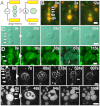
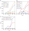
Mechanisms that enabled primitive cell membranes to self-reproduce have been discussed based on the physicochemical properties of fatty acids; however, there must be a transition to modern cell membranes composed of phospholipids [Budin I, Szostak JW (2011) Proc Natl Acad Sci USA 108:5249-5254]. Thus, a growth-division mechanism of membranes that does not depend on the chemical nature of amphiphilic molecules must have existed. Here, we show that giant unilamellar vesicles composed of phospholipids can undergo the coupled process of fusion and budding transformation, which mimics cell growth and division. After gaining excess membrane by electrofusion, giant vesicles spontaneously transform into the budded shape only when they contain macromolecules (polymers) inside their aqueous core. This process is a result of the vesicle maximizing the translational entropy of the encapsulated polymers (depletion volume effect). Because the cell is a lipid membrane bag containing highly concentrated biopolymers, this coupling process that is induced by physical and nonspecific interactions may have a general importance in the self-reproduction of the early cellular compartments.
Conflict of interest statement
The authors declare no conflict of interest.
Figures


Similar articles
-
Giant Polymersome Protocells Dock with Virus Particle Mimics via Multivalent Glycan-Lectin Interactions.Sci Rep. 2016 Aug 31;6:32414. doi: 10.1038/srep32414. Sci Rep. 2016. PMID: 27576579 Free PMC article.
-
Bioadhesive giant vesicles for monitoring hydroperoxidation in lipid membranes.Soft Matter. 2015 Aug 14;11(30):5995-8. doi: 10.1039/c5sm01019e. Epub 2015 Jun 12. Soft Matter. 2015. PMID: 26067909
-
Shape changes of giant liposomes induced by an asymmetric transmembrane distribution of phospholipids.Biophys J. 1992 Feb;61(2):347-57. doi: 10.1016/S0006-3495(92)81841-6. Biophys J. 1992. PMID: 1547324 Free PMC article.
-
Lipid vesicles in pulsed electric fields: Fundamental principles of the membrane response and its biomedical applications.Adv Colloid Interface Sci. 2017 Nov;249:248-271. doi: 10.1016/j.cis.2017.04.016. Epub 2017 Apr 28. Adv Colloid Interface Sci. 2017. PMID: 28499600 Review.
-
Model membrane platforms to study protein-membrane interactions.Mol Membr Biol. 2012 Aug;29(5):144-54. doi: 10.3109/09687688.2012.700490. Epub 2012 Jul 26. Mol Membr Biol. 2012. PMID: 22831167 Review.
Cited by
-
Compartments for Synthetic Cells: Osmotically Assisted Separation of Oil from Double Emulsions in a Microfluidic Chip.Chembiochem. 2019 Oct 15;20(20):2604-2608. doi: 10.1002/cbic.201900152. Epub 2019 Jul 30. Chembiochem. 2019. PMID: 31090995 Free PMC article.
-
L form bacteria growth in low-osmolality medium.Microbiology (Reading). 2019 Aug;165(8):842-851. doi: 10.1099/mic.0.000799. Epub 2019 Apr 8. Microbiology (Reading). 2019. PMID: 30958258 Free PMC article.
-
Asymmetrical Polyhedral Configuration of Giant Vesicles Induced by Orderly Array of Encapsulated Colloidal Particles.PLoS One. 2016 Jan 11;11(1):e0146683. doi: 10.1371/journal.pone.0146683. eCollection 2016. PLoS One. 2016. PMID: 26752650 Free PMC article.
-
L-form bacteria, cell walls and the origins of life.Open Biol. 2013 Jan 8;3(1):120143. doi: 10.1098/rsob.120143. Open Biol. 2013. PMID: 23303308 Free PMC article. Review.
-
Preparation and biomedical applications of artificial cells.Mater Today Bio. 2023 Nov 24;23:100877. doi: 10.1016/j.mtbio.2023.100877. eCollection 2023 Dec. Mater Today Bio. 2023. PMID: 38075249 Free PMC article. Review.
References
-
- Chen IA. GE prize-winning essay. The emergence of cells during the origin of life. Science. 2006;314:1558–1559. - PubMed
-
- Szostak JW, Bartel DP, Luisi PL. Synthesizing life. Nature. 2001;409:387–390. - PubMed
-
- Luisi PL, Ferri F, Stano P. Approaches to semi-synthetic minimal cells: A review. Naturwissenschaften. 2006;93:1–13. - PubMed
-
- Zepik HH, Walde P. Achievements and challenges in generating protocell models. Chembiochem. 2008;9:2771–2772. - PubMed
Publication types
MeSH terms
Substances
LinkOut - more resources
Full Text Sources

