. 2012 Apr 17;109(16):5972-7.
doi: 10.1073/pnas.1108617109. Epub 2012 Apr 2.

Microfluidic integration for automated targeted proteomic assays

Affiliations

Microfluidic integration for automated targeted proteomic assays

Alex J Hughes et al. Proc Natl Acad Sci U S A. .

Abstract

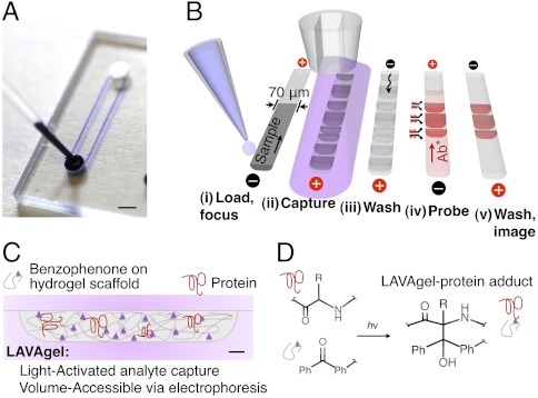
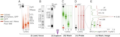
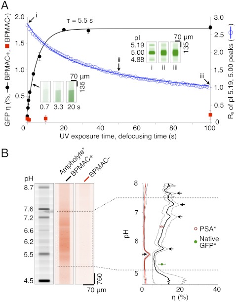
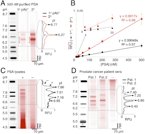
A dearth of protein isoform-based clinical diagnostics currently hinders advances in personalized medicine. A well-organized protein biomarker validation process that includes facile measurement of protein isoforms would accelerate development of effective protein-based diagnostics. Toward scalable protein isoform analysis, we introduce a microfluidic "single-channel, multistage" immunoblotting strategy. The multistep assay performs all immunoblotting steps: separation, immobilization of resolved proteins, antibody probing of immobilized proteins, and all interim wash steps. Programmable, low-dispersion electrophoretic transport obviates the need for pumps and valves. A three-dimensional bulk photoreactive hydrogel eliminates manual blotting. In addition to simplified operation and interfacing, directed electrophoretic transport through our 3D nanoporous reactive hydrogel yields superior performance over the state-of-the-art in enhanced capture efficiency (on par with membrane electroblotting) and sparing consumption of reagents (ca. 1 ng antibody), as supported by empirical and by scaling analyses. We apply our fully integrated microfluidic assay to protein measurements of endogenous prostate specific antigen isoforms in (i) minimally processed human prostate cancer cell lysate (1.1 pg limit of detection) and (ii) crude sera from metastatic prostate cancer patients. The single-instrument functionality establishes a scalable microfluidic framework for high-throughput targeted proteomics, as is relevant to personalized medicine through robust protein biomarker verification, systematic characterization of new antibody probes for functional proteomics, and, more broadly, to characterization of human biospecimen repositories.


Conflict of interest statement

The authors declare no conflict of interest.

Figures

Fig. 1.
Fig. 1.
Design and operation of the microfluidic LAVAgel assay for high-specificity protein isoform analysis. (A) Glass microfluidic device with microchannels linking two fluid reservoirs (dye added for clarity). (Scale bar: 2 mm.) (B) The 80-min five-stage immunoprobing assay is completed in a single microchannel. (C) Schematic of microchannel cross-section depicting principle of the LAVAgel: Analytes are electrophoresed through the reactive nanoporous hydrogel, exposed to UV, and covalently immobilized. (Scale bar: 5 μm.) (D) Schematic of reaction between polypeptide backbone and pendant LAVAgel benzophenone groups. Ph denotes phenyl group. For clarity, the electrophilic triplet state of benzophenone, hydrogen abstraction, and radical intermediates are omitted.
Fig. 2.
Fig. 2.
Characterization of protein isoforms using the single-microchannel 80-min LAVAgel immunoblot. Fluorescence micrographs show (A) loading and IEF of a CE540-labeled protein ladder with 617-nM green WT GFP, and (B) IEF readout via UV excitation. (C) After UV gel photoactivation, the pH gradient is washed out with retention of a portion of each WT GFP isoform. (D and E) Antibody probing of WT GFP with 100 nM Texas red-labeled polyclonal antibody (pAb*) demonstrates specificity and low-background. RFU, relative fluorescence units; CytC, cytochrome C; LCL, lentil lectin; Mb, myoglobin; CA, carbonic anhydrase; BLG, β-lactoglobulin; GOx, glucose oxidase.
Fig. 3.
Fig. 3.
Characterization of LAVAgel photoimmobilization kinetics, capture efficiency, and pH dependence. (A) LAVAgel capture efficiency and resolution losses are optimized by tuning UV exposure duration. Photoactive LAVAgel (BPMAC+, 15 μM WT GFP, ± SD, n = 4, black solid circles) is compared to a nonphotoactive negative control (BPMAC−, red squares). (Inset) Fluorescence micrographs show captured WT GFP fluorescence. Blue open circles and inset images (i–iii) show separation resolution loss for WT GFP isoforms during defocusing. GFP concentration is 617 nM, resolution measured between the pI 5.00 and 5.19 isoforms. (B) Reporter ampholytes (ampholyte*) allow measurement of capture efficiency under focusing conditions for a broad pH range. (Left) Fluorescence micrographs show pI ladder and photocaptured reporter ampholytes after pH gradient washout. (Right) Reporter ampholyte capture efficiency versus pH in BPMA+ and BPMA− LAVAgels, black arrows indicate artifact peaks caused by enhanced local photobleaching of reporter ampholytes in the vicinity of pI marker bands (see SI Text; [ampholyte ∗ ] = 0.025% wt/vol, gray envelopes are ± SD, n = 4).
Fig. 4.
Fig. 4.
LAVAgel assay enables quantitation of PSA isoforms in minimally processed prostate cancer cell lysate and human sera. (A) Fluorescence micrographs and electropherograms for probing of unlabeled PSA purified from human seminal fluid (500 nM): focused pI markers, primary (1°), and secondary (2°) antibody probe signals. Bracketed peak areas used to construct calibration curves. (B) Linear PSA calibration curves for primary (black circles) and secondary (red squares) antibody readouts (RFU, relative fluorescence units; ± SD, n = 4 for all points except 5 nM, n = 2). (C) Primary antibody probing of endogenous PSA isoforms in lysate from a PSA-producing cell line (LAPC-4 cells, +) with negative control lysate (DU145 cells, −). (D) Serum samples from metastatic prostate cancer patients probed with primary antibody to PSA (patients 1 and 2), alongside a low-PSA negative control serum (−).

Similar articles

Cited by

References

    1. Brennan DJ, O’Connor DP, Rexhepaj E, Ponten F, Gallagher WM. Antibody-based proteomics: Fast-tracking molecular diagnostics in oncology. Nat Rev Cancer. 2010;10:605–617. - PubMed
    1. Rifai N, Gillette MA, Carr SA. Protein biomarker discovery and validation: The long and uncertain path to clinical utility. Nat Biotechnol. 2006;24:971–983. - PubMed
    1. Zhu CS, et al. A framework for evaluating biomarkers for early detection: Validation of biomarker panels for ovarian cancer. Cancer Prev Res. 2011;4:375–383. - PMC - PubMed
    1. Esserman L, Shieh Y, Thompson I. Rethinking screening for breast cancer and prostate cancer. JAMA. 2009;302:1685–1692. - PubMed
    1. Etzioni R, et al. The case for early detection. Nat Rev Cancer. 2003;3:243–252. - PubMed

Publication types

LinkOut - more resources