. 2012 Apr 17;109(16):6253-8.
doi: 10.1073/pnas.1119894109. Epub 2012 Apr 2.

Structural and mechanistic analysis of the membrane-embedded glycosyltransferase WaaA required for lipopolysaccharide synthesis

Affiliations

Structural and mechanistic analysis of the membrane-embedded glycosyltransferase WaaA required for lipopolysaccharide synthesis

Helgo Schmidt et al. Proc Natl Acad Sci U S A. .

Abstract

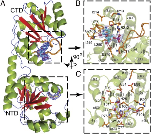
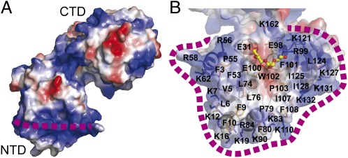
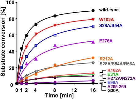
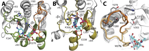
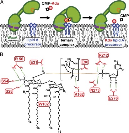
WaaA is a key enzyme in the biosynthesis of LPS, a critical component of the outer envelope of Gram-negative bacteria. Embedded in the cytoplasmic face of the inner membrane, WaaA catalyzes the transfer of 3-deoxy-d-manno-oct-2-ulosonic acid (Kdo) to the lipid A precursor of LPS. Here we present crystal structures of the free and CMP-bound forms of WaaA from Aquifex aeolicus, an ancient Gram-negative hyperthermophile. These structures reveal details of the CMP-binding site and implicate a unique sequence motif (GGS/TX(5)GXNXLE) in Kdo binding. In addition, a cluster of highly conserved amino acid residues was identified which represents the potential membrane-attachment and acceptor-substrate binding site of WaaA. A series of site-directed mutagenesis experiments revealed critical roles for glycine 30 and glutamate 31 in Kdo transfer. Our results provide the structural basis of a critical reaction in LPS biosynthesis and allowed the development of a detailed model of the catalytic mechanism of WaaA.


Conflict of interest statement

The authors declare no conflict of interest.

Figures

Fig. 1.
Fig. 1.
Domain organization of WaaAAAE. (A) Overall structure. β-Strands are shown in red, α-helices in green, and loops and turns in blue. (B) Donor-substrate binding site of the C-terminal domain (CTD). Residues forming the CMP-binding site are shown in orange. Dashed lines indicate hydrogen bonds. In A and B the putative Kdo-binding loop (cβ5–cα4, 263GGTFVNIGGHNLLE276) is highlighted in orange. (C) Putative acceptor-substrate binding site of the N-terminal domain (NTD) occupied by a PEG molecule. Residues potentially involved in acceptor-substrate binding are shown in orange. The final 2|Fo|-|Fc| electron-density maps surrounding the bound CMP and PEG molecules are contoured at 1 σ above the mean. Oxygen atoms are colored red, nitrogen atoms blue, and the phosphorous atom purple. The orientation of B and C with respect to A is indicated by the rotation axis.
Fig. 2.
Fig. 2.
Putative membrane association of WaaAAAE. (A) Surface representation of WaaAAAE calculated with the program ABPS (41), color-coded according to the electrostatic potential (positive potential, blue; negative potential, red). The dashed purple line indicates the horseshoe of basic amino acid residues (for details see Results and Discussion). (B) Transparent surface representation as in A showing the putative membrane-association/acceptor-substrate binding site of the N-terminal domain. Lysine and arginine side chains constituting the horseshoe are shown as blue sticks. The bound PEG molecule is shown in stick representation, demonstrating that the putative membrane-association site and the acceptor-substrate binding site overlap with each other.
Fig. 3.
Fig. 3.
WaaAAAE activity assay. The relative conversion of lipid IVA (a lipid A precursor) to Kdo-lipid IVA is shown for wild-type WaaAAAE and various protein variants at indicated time points. For details see Materials and Methods.
Fig. 4.
Fig. 4.
Comparison of the donor-substrate binding sites of WaaAAAE (A) and a sialyltransferase of the GT-B superfamily [e. g., PM0188 (19)]. (B). Equivalent protein regions involved in the binding of the CMP moiety of the donor substrate are highlighted in green (WaaAAAE) or olive (PM0188). In both structures, an α/β/α motif, overlapping with the α/β/α motif of MurG (16), is involved in CMP binding. Hydrogen bonds are depicted as red dotted lines, and residues involved in CMP binding are shown as sticks. Note that the positions of hydrophobic residues sandwiching the cytosine base (F247/L250 for WaaAAAE and P312/L357 in PM0188) and the glutamate residue contacting the ribose hydroxyls (E276 in WaaAAAE and E338 in PM0188) vary within the equivalent protein regions. The HP motif of PM0188 (H311/P312) overlaps with the highly conserved 211PRH213 triplet of WaaAAAE. The putative Kdo-binding loop of WaaAAAE in A is highlighted in orange. For clarity, the 3F-Neu5Ac moiety of the donor substrate in B has been omitted. (C) Conformational changes upon donor-substrate binding as evident from superimposed structures of PM0188 (23). In the apo form, W270 (orange) is part of a hydrophobic cluster. Binding of the donor-substrate analog CMP-3F-Neu5Ac brings W270 (beige) into contact with the 3F-Neu5Ac moiety of the donor substrate. For clarity, the perspective of C is different from B.
Fig. 5.
Fig. 5.
Conservation and tryptophan-quenching experiments in the putative acceptor-substrate binding site of WaaAAAE. (A) Surface conservation plot generated with Consurf (42) and color-coded as cyan (variable) to white (average conservation) to purple (high conservation). The base of the putative acceptor-substrate binding site harbors several highly conserved amino acid residues such as W102. The PEG molecule is shown in stick representation. (B) Tryptophan-fluorescence difference spectra obtained by subtracting the spectra measured for the isolated individual protein solutions from those recorded in the presence of lipid IVA (a lipid A precursor). An increase in tryptophan fluorescence is observed for the wild-type protein and the E100A variant used as a control but not for the W102A variant, indicating that the hydrophobic part of lipid IVA interacts with W102.
Fig. 6.
Fig. 6.
Schematic representation of the model for Kdo transfer in WaaAAAE. (A) Membrane association and large-scale conformational changes upon substrate binding. The dashed purple line marks the horseshoe-like arrangement of basic residues interacting with the negatively charged groups of membrane components. Upon substrate binding, reorientation of the Kdo-binding loop (orange) and domain closure takes place, followed by the Kdo transfer step. (B) Interactions proposed to play a role in substrate binding and catalysis. Tetraacyl-4′-phosphate lipid A of A. aeolicus is shown as the acceptor for Kdo glycosylation. Galacturonic acid or phosphate (R1) and ester-bound octadecanoic acid (R2) could make up the natural acceptor. Residue E31 serves as the catalytic base, E98 and K162 interact with the oxocarbenium ion-like intermediate of Kdo, and R212 and N273 are involved in phosphate binding. The crucial 4′-phosphate group of the acceptor substrate interacts with R56 and the highly conserved S28 and S54. Hydrogen bonds and electrostatic contacts are drawn as green and brown dashed lines, respectively.

References

    1. Wiese A, Brandenburg K, Ulmer AJ, Seydel U, Müller-Loennies S. The dual role of lipopolysaccharide as effector and target molecule. Biol Chem. 1999;380:767–784. - PubMed
    1. Raetz CR, Whitfield C. Lipopolysaccharide endotoxins. Annu Rev Biochem. 2002;71:635–700. - PMC - PubMed
    1. Holst O. The structures of core regions from enterobacterial lipopolysaccharides - an update. FEMS Microbiol Lett. 2007;271:3–11. - PubMed
    1. Belunis CJ, Clementz T, Carty SM, Raetz CR. Inhibition of lipopolysaccharide biosynthesis and cell growth following inactivation of the kdtA gene in Escherichia coli. J Biol Chem. 1995;270:27646–27652. - PubMed
    1. Rick PD, Young DA. Isolation and characterization of a temperature-sensitive lethal mutant of Salmonella typhimurium that is conditionally defective in 3-deoxy-d-manno-octulosonate-8-phosphate synthesis. J Bacteriol. 1982;150:447–455. - PMC - PubMed

Publication types

MeSH terms

LinkOut - more resources