. 2012 Apr 17;109(16):6229-34.
doi: 10.1073/pnas.1200470109. Epub 2012 Apr 2.

NSP4, an elastase-related protease in human neutrophils with arginine specificity

Affiliations

NSP4, an elastase-related protease in human neutrophils with arginine specificity

Natascha C Perera et al. Proc Natl Acad Sci U S A. .

Abstract

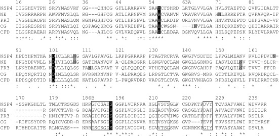
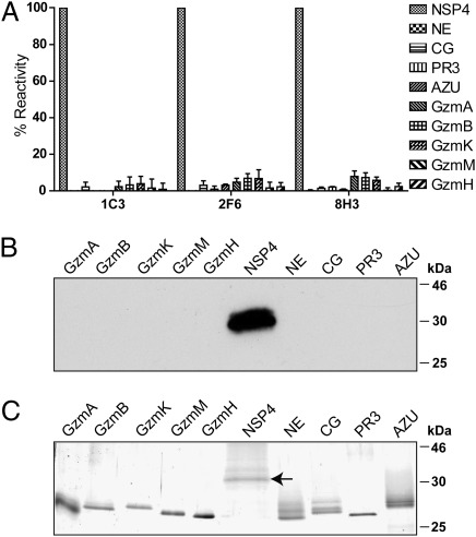
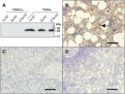
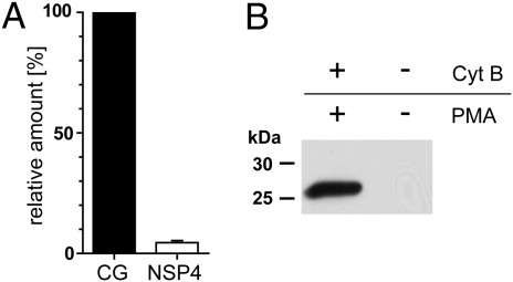
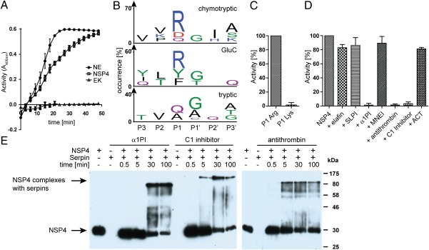
Neutrophil serine proteases (NSPs) in cytoplasmic granules of neutrophils are regarded as important antimicrobial defense weapons after engulfment and exposure of pathogens to the content of primary granules. Despite intensive studies on neutrophils during the last three decades, only three active serine proteases, neutrophil elastase (NE), cathepsin G (CG), and proteinase 3 (PR3) have been identified in these short-lived cells. Here, we report on the identification of a fourth serine protease (NSP4) with 39% identity to NE and PR3, but arginine specificity, yet sharing features like propeptide processing by dipeptidyl peptidase I, storage, and release as an active enzyme with the three active proteases. We established monoclonal antibodies against NSP4, excluded cross-reactivity to human granzymes, NE, CG, PR3, and azurocidin, and screened for NSP4 protein expression in various human tissues and blood leukocyte populations. Only granulocyte precursors and neutrophil populations from peripheral blood were positive. The content of NSP4 in neutrophil lysates, however, was about 20-fold lower compared with CG. Upon neutrophil activation, NSP4 was released into the supernatant. Profiling its specificity with peptide libraries from Escherichia coli revealed a preference for arginine in P1; it cleaved Tyr-Arg-Phe-Arg-AMC and Ala-Pro-Nva-thiobenzyl esters. NSP4 was inhibited by α(1)-proteinase inhibitor (α(1)-antitrypsin), C1 inhibitor, and most efficiently by antithrombin-heparin, but not by elafin, secretory leukocyte protease inhibitor, α(1)-antichymotrypsin, and monocyte-neutrophil elastase inhibitor. Functional specialization and preferred natural substrates of NSP4 remain to be determined to understand the biological interplay of all four NSPs during neutrophil responses.


Conflict of interest statement

The authors declare no conflict of interest.

Figures

Fig. 1.
Fig. 1.
Sequence comparison between all four catalytically active human neutrophil serine proteases and complement factor D. Topologically aligned amino acid residues (single letter code) are numbered according to chymotrypsinogen. The catalytic triad of His57, Asp102, and Ser195 is highlighted in black. NSP4 carries two N-linked glycosylation sites marked in gray. Residues that shape the substrate binding pocket in NE are boxed (189–195, 213–216, 226–228) for all proteases and are similar between NSP4, NE, and PR3. An asterisk (*) below the alignments indicates positions which agree completely; a colon (:) or a period (.) indicate conservation of residues with strong or weak similarities (scoring > 0.5 or = < 0.5 in the Gonnet PAM 250 matrix), respectively.
Fig. 2.
Fig. 2.
Identification of NSP4 specific rat monoclonal antibodies. (A) Only 3 of 14 monoclonal antibodies obtained from hybridomas after immunization of rats, named mAb 1C3, 2F6, and 8H3, were specific for NSP4. NSP4, the human Gzms (A, B, K, M, and H), and all known human NSPs were coated on microtiter plates. Supernatants of hybridoma clones secreting rat antibodies were analyzed in an ELISA using a secondary anti-rat HRP-conjugated polyclonal Ab. Percent reactivity was calculated by setting the mean reactivity of mAbs with NSP4 to 100%. Data are the mean from three independent experiments (n = 3, ± SD). (B) NSP4, the five human Gzms and human NSPs were separated by SDS/PAGE, transferred to Hybond ECL transfer membrane, and then probed with rat anti-NSP4 mAb 1C3, 2F6, or 8H3 and secondary HRP-labeled Ab. (C) Similar amounts of all tested proteins used as in A and B are shown after reducing SDS/PAGE and subsequent silver-staining. A horizontal arrow points to the glycosylated NSP4, carrying an amino-terminal extension (i.e., S-tag and an enterokinase cleavage peptide sequence).
Fig. 3.
Fig. 3.
NSP4 is present in granulocytes. (A) Total cell lysates of peripheral blood mononuclear cells (PBMCs) and PMNs, which are almost exclusively neutrophil granulocytes, were analyzed by Western blotting using anti-NSP4 mAbs and secondary anti-rat HRP-labeled Ab. Natural NSP4 of PMNs runs lower than recombinant S-tag-NSP4 (Fig. 2B) because it is N-terminally processed and has shorter carbohydrate chains. (B–D) Staining of different human tissue samples using anti-NSP4 mAbs and the Ultravision LP staining kit. NSP4 was detected in neutrophil granulocytes and precursors in bone marrow tissue (arrow, B). NSP4 was not detected in lymph node (C) or spleen tissues (D). (Scale bars, 100 μm.)
Fig. 4.
Fig. 4.
Total amount of NSP4 in PMNs is 20-fold lower than that of CG; nevertheless, NSP4 is detected in supernatants of stimulated PMNs. (A) NSP4 and CG in total cell lysates of PMNs were compared with known amounts of purified CG and NSP4 by semiquantitative Western blotting using monospecific anti-CG antibodies and anti-NSP4 mAbs, respectively. Content of NSP4 in PMNs is given as a percentage of CG present in PMNs. Data were pooled from three independent experiments (n = 3, ± SD). (B) PMNs were incubated at 37 °C in the presence or absence of 5 μg/mL cytochalasin B (Cyt B) and 200 ng/mL PMA for 30 min. Cell-free supernatants were analyzed by Western blotting with anti-NSP4 mAbs.
Fig. 5.
Fig. 5.
NSP4 preferably cleaves after arginine residues and is inhibited by α1PI, C1 inhibitor, and antithrombin. (A) After removing the propeptide extension with enterokinase (EK), proteolytic activity of recombinant NSP4 was measured using the thiobenzyl ester substrate Boc-Ala-Pro-Nva-4-chloro-SBzl and DTNB. NSP4 was used at a concentration of 3 × 10⁻⁶ M. NE and the convertase EK served as positive and negative controls at concentrations of 1 × 10⁻⁷ M and 3 × 10⁻² U/μL, respectively. Substrate cleavage was determined by absorbance measurements at 405 nm. Data are results from three experiments, ± SD. (B) PICS specificity profile for NSP4 with chymotrypsin, GluC, and trypsin libraries generated from E. coli. Sequence logos were generated with IceLogo (26). (C) Enzymatic activity of 0.25 μM NSP4 was measured using AMC substrates with either Arg or Lys in P1 [H-Tyr-Arg-Phe-(Arg/Lys)-AMC] at 1 mM substrate concentration. Percent activity was calculated by setting the mean activity of NSP4 with P1 Arg to 100%. Data are the mean from three independent experiments (n = 3, ± SD). (D) 0.25 μM NSP4 was preincubated with 10 μM elafin, 10 μM SLPI, 25 μM α1PI, 3.3 μM MNEI with 1 mM DTT, 5.6 μM antithrombin with 300 U/mL heparin, 2.5 μM C1 inhibitor, or 5.6 μM ACT. Proteolytic activity was determined using H-Tyr-Arg-Phe-Arg-AMC. Percent activity was calculated by setting the mean activity of NSP4 without inhibitor to 100%. Data are the mean from three independent experiments (n = 3, ± SD), except for MNEI and ACT (n = 2, ± SD). (E) 0.8 μM NSP4 was incubated at 37 °C with 40 μM α1PI, 4 μM C1 Inhibitor or 5 μM antithrombin with 300 U/mL heparin and analyzed after different timepoints by Western blotting with anti NSP4 mAbs. For comparisons, NSP4 and the serpins alone were treated similarly.

References

    1. Belaaouaj A. Neutrophil elastase-mediated killing of bacteria: Lessons from targeted mutagenesis. Microbes Infect. 2002;4:1259–1264. - PubMed
    1. Segal AW. How neutrophils kill microbes. Annu Rev Immunol. 2005;23:197–223. - PMC - PubMed
    1. Williams R. Killing controversy. J Exp Med. 2006;203:2404. - PMC - PubMed
    1. Weiss SJ. Tissue destruction by neutrophils. N Engl J Med. 1989;320:365–376. - PubMed
    1. Kessenbrock K, Dau T, Jenne DE. Tailor-made inflammation: How neutrophil serine proteases modulate the inflammatory response. J Mol Med (Berl) 2011;89:23–28. - PMC - PubMed

Publication types

MeSH terms