. 2012 Apr 17;109(16):6130-5.
doi: 10.1073/pnas.1201351109. Epub 2012 Apr 2.

Inducible NOS-induced chloride intracellular channel 4 (CLIC4) nuclear translocation regulates macrophage deactivation

Affiliations

Inducible NOS-induced chloride intracellular channel 4 (CLIC4) nuclear translocation regulates macrophage deactivation

Mariam Malik et al. Proc Natl Acad Sci U S A. .

Abstract

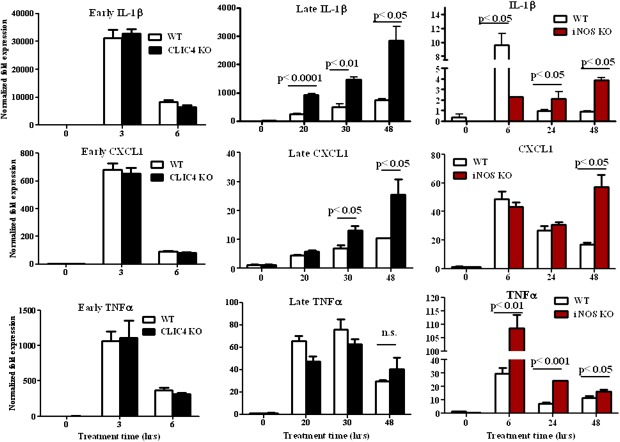
Nuclear translocation of cytosolic CLIC4 is an essential feature of its proapoptotic and prodifferentiation functions. Here we demonstrate that CLIC4 is induced concurrently with inducible nitric oxide synthase (iNOS) and S-nitrosylated in proinflammatory peritoneal macrophages. Chemical inhibition or genetic ablation of iNOS inhibits S-nitrosylation and nuclear translocation of CLIC4. In macrophages, iNOS-induced nuclear CLIC4 coincides with the pro- to anti-inflammatory transition of the cells because IL-1β and CXCL1 mRNA remain elevated in CLIC4 and iNOS knockout macrophages at late time points, whereas TNFα mRNA is elevated only in the iNOS knockout macrophages. Active IL-1β remains elevated in CLIC4 knockout macrophages and in macrophages in which CLIC4 nuclear translocation is prevented by the NOS inhibitor l-NAME. Moreover, overexpression of nuclear-targeted CLIC4 down-regulates IL-1β in stimulated macrophages. In mice, genetically null for CLIC4, the number of phagocytosing macrophages stimulated by LPS is reduced. Thus, iNOS-induced nuclear CLIC4 is an essential part of the macrophage deactivation program.


Conflict of interest statement

The authors declare no conflict of interest.

Figures

Fig. 1.
Fig. 1.
CLIC4 is highly expressed in primary macrophages (Mϕ), is S-nitrosylated, and translocates to the nucleus in response to LPS/IFNγ. (A and B) RNA from mouse hematopoietic cells (A) or mouse macrophages (B), stimulated or unstimulated, as indicated, was isolated and used for CLIC4 and iNOS real-time PCR analysis. In A, RAW and macrophages were stimulated for 6 h with LPS/IFNγ. Bars represent the mean ± SEM of three replicates. Statistical significance was determined using a two-tailed unpaired Student's t test and is indicated. (C) Whole-cell lysates from stimulated or unstimulated macrophages were used for immunoblotting with CLIC4, CLIC1, and iNOS proteins. (D and E) Primary mouse macrophages were treated with LPS and IFNγ (1 μg/mL and 10 ng/mL, respectively) for 18 h. (D) Lysates were used to perform biotin switch assays to detect S-nitrosylation. Lysate from LPS/IFNγ-treated cells was also used for a reaction that omitted ascorbate (“no Asc.”). Five percent of lysates were used as input controls. Assays were immunoblotted for CLIC4. Lane 2 of CLIC4 input is representative for both LPS/IFNγ-treated pull-down assays. The SNO-CLIC4:CLIC4 input ratio was calculated for all treatments across four independent experiments. Statistical significance was determined using a two-tailed unpaired Student's t test. Control versus LPS/IFN treatment has a P < 0.01 and LPS/IFN versus LPS/IFN (no ascorbate) has a P < 0.05. (E) Macrophage cells were immunostained for CLIC4 and visualized with confocal microscopy. Inset nuclei are visualized using DAPI.
Fig. 2.
Fig. 2.
Pharmacologic inhibition or genetic ablation of iNOS inhibits S-nitrosylation and nuclear translocation of CLIC4 in murine macrophages. (AC) RAW 264.7 macrophages were pretreated with (N-(3-(Aminomethyl) benzyl) acetamidine dihydrochloride) 1400 W (100 μM) for 1 h in indicated cases before treatment with LPS and IFNγ (1 μg/mL and 10 ng/mL, respectively) for 24 h. (A) Lysates were used to perform biotin switch assays. Lysate from LPS/IFNγ-treated cells was also used for a reaction that omitted biotin. Lane 3 of CLIC4 input is representative for both LPS/IFNγ-treated pull-down assays. Five percent of lysates were used as input controls and immunoblotted for CLIC4. SNO-CLIC4:CLIC4 input ratio was calculated for treatments across three independent experiments. Statistical significance was determined using a two-tailed unpaired Student's t test. 1400W versus LPS/IFN treatment has a P < 0.05 and LPS/IFN versus LPS/IFN +1400W has a P < 0.05. (B) Media from treated plates was collected and assayed for nitrite + nitrate levels in control and treated cells. (C) Cells were immunostained for CLIC4, and nuclei were stained with DAPI and visualized with confocal microscopy. (DF) Primary mouse macrophages from iNOS knockout and wild-type mice were treated with LPS and IFNγ (1 μg/mL and 10 ng/mL, respectively) for 18 h. (D) Lysates were used to perform biotin switch assays. Lysate from LPS/IFNγ-treated wild-type cells was also used for a reaction that omitted ascorbate (“no Asc.”). Five percent of lysates were used as input controls and immunoblotted for CLIC4. (E) Cells were used for subcellular fractionation, and nuclear and cytosolic lysates were immunoblotted for CLIC4 and β-actin. (F) Macrophage cells were immunostained for CLIC4 and visualized with confocal microscopy. Nuclei were counterstained with DAPI and are pseudocolored red for better visualization.
Fig. 5.
Fig. 5.
Chemical inhibition of iNOS activity enhances IL-1β and iNOS levels, whereas overexpression of CLIC4 down-regulates IL-1β and iNOS. (AC) Primary mouse macrophages from wild-type mice were treated with LPS and IFNγ (1 μg/mL and 10 ng/mL, respectively) for 24 h (A) or for 48 h (B and C). Cells were pretreated with 2.5 mM l-NAME for 1 h where indicated. (A) Nuclear and cytosolic lysates were prepared and immunoblotted for CLIC4 and β-actin antibodies. (B) Whole-cell lysates were immunoblotted for IL-1β, iNOS, CLIC4, CLIC1, and β-actin. (C) Media from treated samples was assayed for cleaved IL-1β by ELISA. Bars represent the mean ± SEM of three replicates. (D) RAW macrophages were transduced with adenoviruses expressing nuclear-targeted CLIC4 (NUC-CLIC4), HA-tagged CLIC4 (HA-CLIC4), or GFP proteins overnight followed by treatment with LPS and IFNγ (1 μg/mL and 10ng/mL, respectively) for 24 h. Whole-cell lysates were immunoblotted for IL-1β, iNOS, CLIC4, GFP, and β-actin.
Fig. 3.
Fig. 3.
CLIC4 and iNOS knockout macrophages exhibit similar dysregulation of IL-1β and CXCL1 but not TNFα mRNA after prolonged stimulation with LPS/IFNγ. Primary mouse macrophages from CLIC4 knockout, iNOS knockout, and wild-type mice were treated with LPS and IFNγ (1 μg/mL and 10 ng/mL, respectively) for the indicated times. Lysates were used for real-time PCR analysis of IL-1β, CXCL1, and TNFα mRNA. Bars represent the mean ± SEM of three replicates. Statistical significance was determined using a two-tailed unpaired Student's t test and is indicated.
Fig. 4.
Fig. 4.
CLIC4 knockout macrophages show enhanced levels of IL-1β activity, lower levels of F4/80 receptor, and reduced numbers of phagocytosing macrophages in vivo. (A and B) Primary mouse macrophages from CLIC4 knockout and wild-type mice were treated with LPS and IFNγ (1 μg/mL and 10 ng/mL, respectively) for the indicated times. (A) Whole-cell lysates were immunoblotted for CLIC4, IL-1β, iNOS, and β-actin, as indicated. (B) Media from quadruplicate samples was assayed for cleaved IL-1β by ELISA. Bars represent the mean ± SEM. (C) Wild-type and CLIC4 knockout animals were injected with thioglycollate and 6 d later injected with LPS (12.5 μg/g) for 24 h. Animals were injected with pHrodo-labeled E. coli for 1 h before peritoneal macrophages were collected by lavage and stained for APC-F4/80 and with DAPI. Viable cells from each animal were analyzed for F4/80 and pHrodo fluorescence using flow cytometry. Statistical significance was determined using a two-tailed unpaired Student's t test and is indicated. Each dot represents results from a single animal.

References

    1. Qian Z, Okuhara D, Abe MK, Rosner MR. Molecular cloning and characterization of a mitogen-activated protein kinase-associated intracellular chloride channel. J Biol Chem. 1999;274:1621–1627. - PubMed
    1. Tulk BM, Kapadia S, Edwards JC. CLIC1 inserts from the aqueous phase into phospholipid membranes, where it functions as an anion channel. Am J Physiol Cell Physiol. 2002;282:C1103–C1112. - PubMed
    1. Berryman M, Bruno J, Price J, Edwards JC. CLIC-5A functions as a chloride channel in vitro and associates with the cortical actin cytoskeleton in vitro and in vivo. J Biol Chem. 2004;279:34794–34801. - PubMed
    1. Fernández-Salas E, et al. mtCLIC/CLIC4, an organellular chloride channel protein, is increased by DNA damage and participates in the apoptotic response to p53. Mol Cell Biol. 2002;22:3610–3620. - PMC - PubMed
    1. Shiio Y, et al. Quantitative proteomic analysis of myc-induced apoptosis: A direct role for Myc induction of the mitochondrial chloride ion channel, mtCLIC/CLIC4. J Biol Chem. 2006;281:2750–2756. - PubMed

Publication types

MeSH terms

LinkOut - more resources