. 2012 Apr 17;109(16):6193-8.
doi: 10.1073/pnas.1117490109. Epub 2012 Apr 2.

Preventing bacterial DNA release and absent in melanoma 2 inflammasome activation by a Legionella effector functioning in membrane trafficking

Affiliations

Preventing bacterial DNA release and absent in melanoma 2 inflammasome activation by a Legionella effector functioning in membrane trafficking

Jianning Ge et al. Proc Natl Acad Sci U S A. .

Abstract

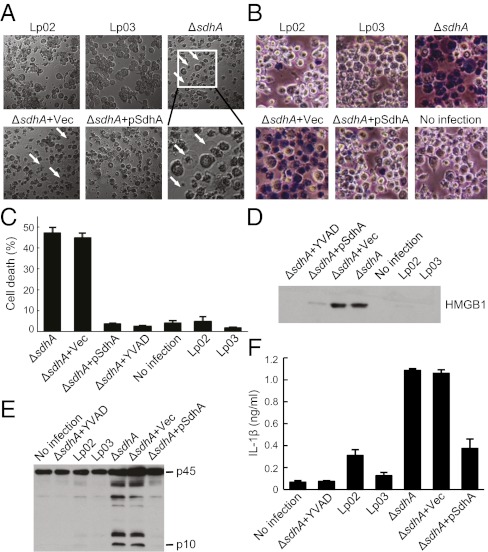
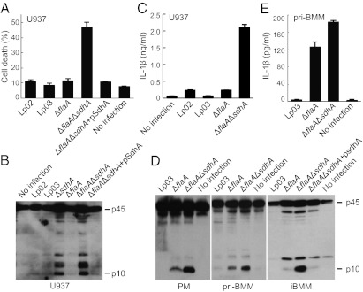
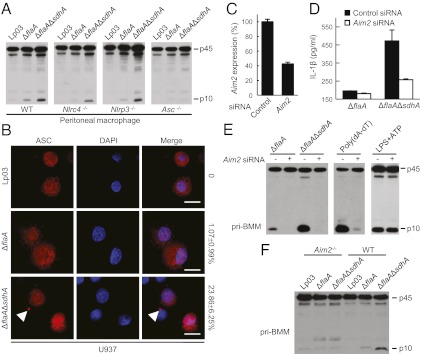
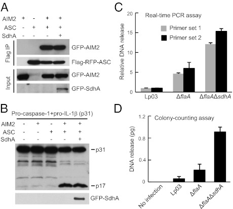
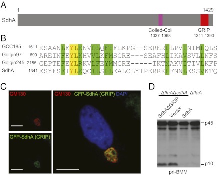
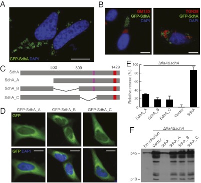
Legionella pneumophila, the causative agent of Legionnaires' pneumonia, resides in a distinct vacuole structure called Legionella-containing vacuole (LCV). The LCV resists fusion with the lysosome and permits efficient bacterial replication in host macrophages, which requires a Dot/Icm type IVB secretion system. Dot/Icm-translocated effector SdhA is critical for L. pneumophila intracellular growth and functions to prevent host cell death. Here, we show that the absence of SdhA resulted in elevated caspase-1 activation and IL-1β secretion as well as macrophage pyroptosis during Legionella infection. These inflammasome activation phenotypes were independent of the established flagellin-NAIP5-NLRC4 axis, but relied on the DNA-sensing AIM2 inflammasome. We further demonstrate that Legionella DNA was released into macrophage cytosol, and this effect was significantly exaggerated by the absence of SdhA. SdhA bears a functional Golgi-targeting GRIP domain that is required for preventing AIM2 inflammasome activation. Ectopically expressed SdhA formed a unique ring-shape membrane structure, further indicating a role in membrane trafficking and maintaining LCV membrane integrity. Our data together suggest a possible link, mediated by the function of SdhA, between LCV trafficking/maturation and suppression of host innate immune detection.


Conflict of interest statement

The authors declare no conflict of interest.

Figures

Fig. 1.
Fig. 1.
Deletion of sdhA triggers pyroptosis and caspase-1 activation in U937 cells. (A and B) Morphological examination of effects of sdhA deletion during L. pneumophila infection of PMA-differentiated U937 cells (MOI = 10). Lp02 is used as the wild-type strain; Lp03 is a dotA mutant. pSdhA is a complementation plasmid expressing SdhA. Differential interference contrast images (5 h after infection) are shown with cells stained with Trypan blue in B. Arrows indicate pyroptotic cells in A. (C and D) Lactate dehydrogenase (LDH) (C) and HMGB1 (D) release assays of effects of sdhA deletion. Shown in C are percentages of cell death as mean values ± SD (error bars) from four independent experiments. Anti-HMGB1 immunoblot of culture media is shown in D. YVAD, a caspase-1 inhibitor. (E and F) Caspase-1 activation and IL-1β release assays of sdhA deletion. Shown in E is anti–caspase-1 immunoblot of culture supernatants. p45, procaspase-1; p10, the processed mature form of caspase-1. IL-1β ELISA data shown in F are as mean values ± SD (error bars) from three independent experiments.
Fig. 2.
Fig. 2.
ΔsdhA-induced inflammasome activation is flagellin-independent. U937 cells were used for infection in AC and mouse macrophages were assayed in D and E. LDH release in A shows percentages of cell death as mean values ± SD (error bars) from four independent experiments. Shown in B and D are anti–caspase-1 immunoblots of culture supernatants. IL-1β ELISA data shown in C and E are mean values ± SD (error bars) from three independent experiments. iBMM, immortalized bone marrow macrophages (C57/BL6 background); PM, peritoneal macrophages (129S background); pri-BMM, primary bone marrow macrophages (129S background). Culture supernatants collected from 2 to 6 h after infection were subjected to analyses in D and E.
Fig. 3.
Fig. 3.
ΔsdhA-induced inflammasome activation requires Aim2 and Asc. (A) ΔsdhA-induced caspase-1 activation in Nlrc4−/−, Nlrp3−/−, and Asc−/− macrophages. Peritoneal macrophages prepared from C57/BL6 (WT) or indicated knockout mice were infected with indicated L. pneumophila strains. (B) ASC foci formation in U937 cells infected with sdhA+ or sdhA strains (MOI = 10). Statistics of percentages of cells showing the ASC pyroptosome (arrowhead) are listed on the right to the merged confocal images. (Scale bars: 7.5 μm.) (CE) Effects of Aim2 knockdown on ΔsdhA-induced inflammasome activation. BMMs from 129S mice were transfected with Aim2 targeting or a control siRNA before indicated infections. qRT-PCR measurements of knockdown efficiency (C) are shown as mean values ± SEM (error bars) from three independent experiments. The supernatants were subjected to IL-1β ELISA (D) or anti–caspase-1 immunoblotting (E). Mean values ± SEM (error bars) from two independent experiments are shown in D. (F) ΔsdhA-induced caspase-1 activation in Aim2−/− macrophages. Peritoneal macrophages prepared from C57/BL6 (WT) or Aim2−/− mice were infected with indicated L. pneumophila strains.
Fig. 4.
Fig. 4.
SdhA inhibits Legionella DNA release into macrophage cytosol. (A and B) SdhA does not directly target the AIM2–ASC–caspase-1 pathway. 293T cells transfected with indicated plasmid combination were subjected to anti-Flag immunoprecipitation and/or immunoblotting. p17, the processed mature form of IL-1β. (C and D) Bacterial DNA release into macrophage cytosol during infection and effects of sdhA deletion. PMA-differentiated U937 cells were infected with indicated L. pneumophila strains harboring a reporter plasmid. DNA fractions extracted from infected macrophage cytosol were subjected to qRT-PCR analysis (C) or colony-counting assay (D) to measure relative plasmid DNA release (detailed in SI Materials and Methods). Mean values ± SEM (error bars) from three independent experiments are shown.
Fig. 5.
Fig. 5.
SdhA harbors a Golgi-targeting GRIP domain that is required for inhibiting inflammasome. (A) The domain structure of SdhA. (B) Sequence alignment of residues 1341–1381 in SdhA with GRIP domains from several mammalian golgins. Similar residues are highlighted in green, and identical ones are in yellow. (C) SdhA GRIP domain localization in HeLa cells. The Golgi was visualized by GM130 staining. Representative confocal immunofluorescence images are shown. (Scale bars: 7.5 μm.) (D) Complementation of ΔflaAΔsdhA strain by plasmid-encoded SdhA or the SdhAΔGRIP mutant. BMMs from 129S mice were infected with L. pneumophila deletion or rescue strains as indicated. Caspase-1 immunoblot of culture supernatant is shown.
Fig. 6.
Fig. 6.
SdhA forms a distinct ring-shape membrane structure and mutants deficient in ring formation fail to complement ΔsdhA strain. (A and B) GFP-SdhA forms a ring-shape membrane structure that is negative for Golgi markers. GM130 and TGN38 antibodies stain the Golgi structure. (Scale bars: 7.5 μm.) (C) A schematic drawing of SdhA truncation mutant assayed in (DF). SdhA_A, Δ1–499aa; SdhA_B, Δ501–809aa; SdhA_C, Δ847–1104aa. (D) Localization of GFP-tagged SdhA truncation mutants in HeLa cells. (Scale bars: 7.5 μm.) (E and F) Complementation assays of SdhA truncation mutants. PMA-differentiated U937 cells were infected with ΔflaAΔsdhA strain transformed with a plasmid expressing SdhA or the indicated truncation mutants. Cell death measured by LDH release was assayed in E, and rescue effects relative to vector-complemented strain are shown as mean values ± SD (error bars) from three independent experiments.

References

    1. Isberg RR, O'Connor TJ, Heidtman M. The Legionella pneumophila replication vacuole: Making a cosy niche inside host cells. Nat Rev Microbiol. 2009;7:13–24. - PMC - PubMed
    1. Marra A, Blander SJ, Horwitz MA, Shuman HA. Identification of a Legionella pneumophila locus required for intracellular multiplication in human macrophages. Proc Natl Acad Sci USA. 1992;89:9607–9611. - PMC - PubMed
    1. Berger KH, Isberg RR. Two distinct defects in intracellular growth complemented by a single genetic locus in Legionella pneumophila. Mol Microbiol. 1993;7:7–19. - PubMed
    1. Vogel JP, Andrews HL, Wong SK, Isberg RR. Conjugative transfer by the virulence system of Legionella pneumophila. Science. 1998;279:873–876. - PubMed
    1. Hubber A, Roy CR. Modulation of host cell function by Legionella pneumophila type IV effectors. Annu Rev Cell Dev Biol. 2010;26:261–283. - PubMed

Publication types

MeSH terms

LinkOut - more resources