Review
. 2012:2012:481840.
doi: 10.1155/2012/481840. Epub 2012 Feb 28.

Endothelial dysfunction in diabetes mellitus: possible involvement of endoplasmic reticulum stress?

Affiliations
Review

Endothelial dysfunction in diabetes mellitus: possible involvement of endoplasmic reticulum stress?

Basma Basha et al. Exp Diabetes Res. 2012.

Abstract

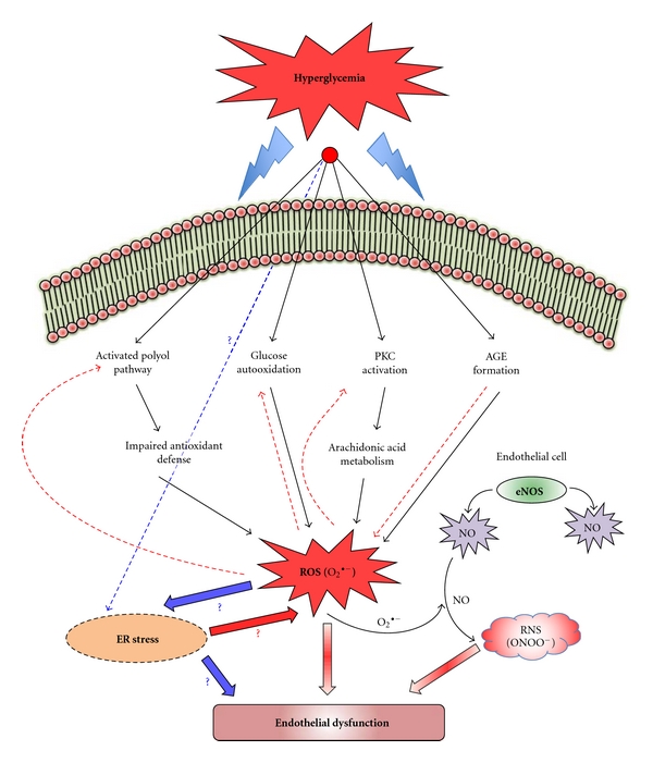
The vascular complications of diabetes mellitus impose a huge burden on the management of this disease. The higher incidence of cardiovascular complications and the unfavorable prognosis among diabetic individuals who develop such complications have been correlated to the hyperglycemia-induced oxidative stress and associated endothelial dysfunction. Although antioxidants may be considered as effective therapeutic agents to relieve oxidative stress and protect the endothelium, recent clinical trials involving these agents have shown limited therapeutic efficacy in this regard. In the recent past experimental evidence suggest that endoplasmic reticulum (ER) stress in the endothelial cells might be an important contributor to diabetes-related vascular complications. The current paper contemplates the possibility of the involvement of ER stress in endothelial dysfunction and diabetes-associated vascular complications.


Figures

Figure 1
Figure 1
Hyperglycemia-induced oxidative stress and endothelial dysfunction (possible role of ER stress). High glucose levels in circulation can divert glucose into alternative biochemical pathways leading to the increase in advanced glycation end products (AGEs), glucose autooxidation, hexosamine and polyol flux, and activation of classical isoforms of protein kinase C, that are considered to be the mediators of hyperglycemia-induced cellular injury. Many different pathways involved in hyperglycemia-mediated endothelial dysfunction induced by hyperglycemia lead to considerable generation of reactive oxygen species (ROS), which is responsible for the oxidative stress. The excessive ROS so formed can then aggravate cellular injury by promoting activation of the biochemical pathways (red dotted arrows) that initiate ROS generation in the first place as a response to hyperglycemia, thus completing a vicious cycle. Superoxide anion (O2 ∙−) can also react with NO to yield peroxynitrite which is also known to be a mediator of endothelial dysfunction. It is still unclear whether the ER stress response can be initiated as a direct response to the increasing load on protein synthesis and maturation due to hyperglycemia or due to the hyperglycemia-associated oxidative stress. The possibility that ER stress response also can lead to excessive ROS formation cannot be ruled out.
Figure 2
Figure 2
Signal transduction events associated with ER stress. The accumulation of misfolded proteins and disruption of Ca2+ homeostasis in the ER disrupt ER function leading to ER stress. The unfolded protein response (UPR) is initiated as a response to this stress, where GRP78, PERK, IRE1, and ATF6 play a central role. However, there might be more unknown mediators of ER stress. The cell initially tries to resolve the ER stress and restore normal cell function by halting protein synthesis and activation of several ER Stress Response (ERSR) adaptation genes, which include chaperones and proteins of the ER-associated degradation (ERAD) system. However, prolonged ER stress leads to the activation of the “alarm response”, leading to cell damage, dysfunction, and finally apoptosis.
Figure 3
Figure 3
Signaling by PERK, IRE1, and ATF6. In an unstressed condition, GRP78, the primary sensor of ER stress binds to transmembrane ER proteins, PERK, IRE1, and ATF6, preventing their activation. The accumulation of unfolded proteins leads to dissociation of GRP78 from PERK, IRE1, and ATF6. GRP78, however, binds to the unfolded protein. GRP78 dissociation leads to PERK and IRE1 oligomerization and trans-autophosphorylation of their cytosolic domains. The active phosphorylated PERK (p-PERK, active) in turn phosphorylates eIF2α (p-eIF2α, inactive), leading to attenuation of global protein synthesis. Under these conditions, selected mRNAs, such as ATF4, are translated, which induces the expression of genes involved in restoring ER homeostasis. ATF4 can activate GADD34, which recruits a phosphatase to dephosphorylate p-PERK and reverse translational attenuation. ER-resident p58IPKs is an inhibitor of PERK. Phosphorylation of IRE1 (p-IRE1) activates its endoribonucleolytic activity, which then excises an intron of the XBP1 mRNA to generate a mature XBP1 mRNA, which then encodes for the active protein, XBP1. The XBP1 protein then translocates into the nucleus and supports the ER stress response. Interaction of p-IRE1 with TRAF2 can elicit the activation of the JNK pathway and caspases leading to apoptosis. GRP78 release from the dimeric ATF6 leads to the translocation of its monomeric form to the Golgi, where it undergoes proteolytic processing by the proteases (S1P and S2P). The cleaved ATF6 then translocates to the nucleus where it activates the ERSR genes and chaperones. Alternatively, CHOP/GADD153 can be activated by ATF4, ATF6, or XBP1, which promotes apoptosis by decreasing the levels of antiapoptotic Bcl-2 in the cell. The dotted arrows denote translocation. XBP1 (a) denotes the mature XBP1 mRNA, while XBP1p denotes the protein product of XBP1 (a). The “+” sign signifies activation.

Similar articles

Cited by

References

    1. World Health Organization, Bode CD. Media Centre-Fact Sheets, Diabetes, 2011, http://www.who.int/mediacentre/factsheets/fs312/en/index.html.
    1. Schalkwijk CG, Stehouwer CDA. Vascular complications in diabetes mellitus: the role of endothelial dysfunction. Clinical Science. 2005;109(2):143–159. - PubMed
    1. Fowler MJ. Microvascular and macrovascular complications of diabetes. Clinical Diabetes. 2008;26(2):77–82.
    1. Grundy SM, Howard B, Smith S, Jr., Eckel R, Redberg R, Bonow RO. Prevention conference VI: diabetes and cardiovascular disease—executive summary: conference proceeding for healthcare professionals from a special writing group of the American Heart Association. Circulation. 2002;105(18):2231–2239. - PubMed
    1. Bakker W, Eringa EC, Sipkema P, Van Hinsbergh VWM. Endothelial dysfunction and diabetes: roles of hyperglycemia, impaired insulin signaling and obesity. Cell and Tissue Research. 2009;335(1):165–189. - PubMed

Publication types

MeSH terms