

Review
doi: 10.1155/2012/548732.
Epub 2012 Feb 26.
Emerging pharmacotherapies for diabetic macular edema
Affiliations
- PMID: 22474425
- PMCID: PMC3299388
- DOI: 10.1155/2012/548732
Item in Clipboard
Review
Emerging pharmacotherapies for diabetic macular edema
Exp Diabetes Res.
2012.
Abstract
Diabetic macular edema (DME) remains an important cause of visual loss in patients with diabetes mellitus. Although photocoagulation and intensive control of systemic metabolic factors have been reported to achieve improved outcomes in large randomized clinical trials (RCTs), some patients with DME continue to lose vision despite treatment. Pharmacotherapies for DME include locally and systemically administered agents. We review several agents that have been studied for the treatment of DME.
Figures
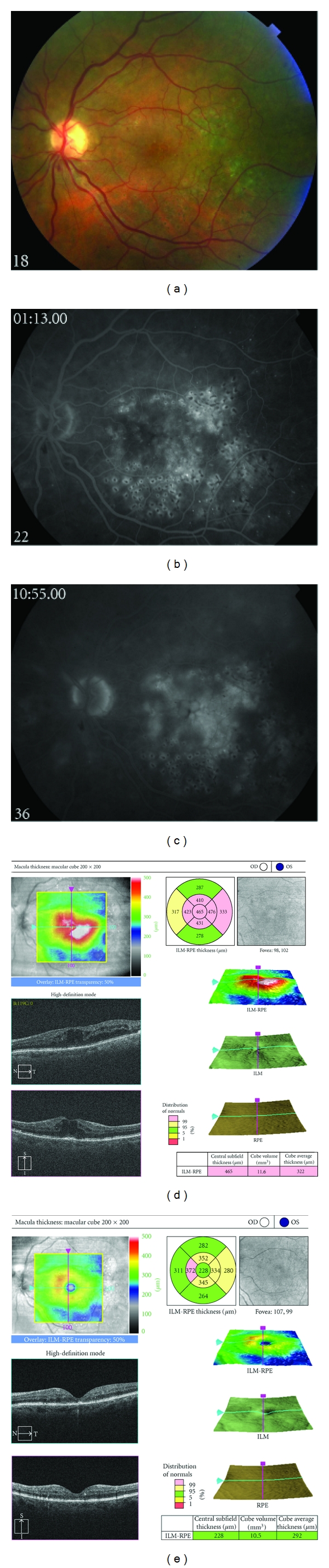
(a) Fundus photograph, left eye, of a patient with persistent diabetic macular edema following focal/grid photocoagulation. (b) Early phase fluorescein angiograph, left eye, demonstrating abnormal hyperfluorescence in the macula. (c) Late phase fluorescein angiograph, left eye, demonstrating profuse leakage consistent with angiographic macular edema. (d) Spectral domain optical coherence tomograph, left eye, demonstrating cystoid macular edema. (e) Following treatment with intravitreal triamcinolone acetonide, 4 mg in 0.1 mL, spectral domain optical coherence tomography demonstrates marked improvement in cystoid macular edema.
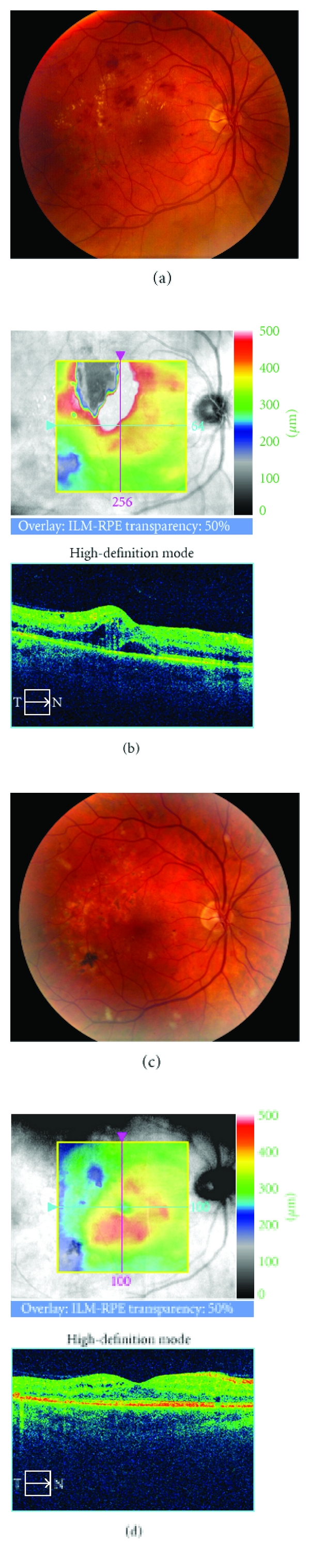
(a) Fundus photograph, right eye, of a patient with persistent diabetic macular edema following focal/grid photocoagulation. (b) Spectral domain optical coherence tomograph, right eye, demonstrates cystoid macular edema and subretinal fluid. (c) Following additional focal/grid photocoagulation and treatment with intravitreal bevacizumab, 1.25 mg in 0.1 mL, fundus photography demonstrates marked improvement in diabetic macular edema. (d) Follow-up spectral domain optical coherence tomography demonstrates marked improvement in intraretinal and subretinal fluid.
Similar articles
-
Emerging pharmacotherapies for diabetic macular edema.Expert Opin Emerg Drugs. 2007 Nov;12(4):591-603. doi: 10.1517/14728214.12.4.591. Expert Opin Emerg Drugs. 2007. PMID: 17979601 Review.
-
Current intravitreal pharmacologic therapies for diabetic macular edema.Postgrad Med. 2015 Aug;127(6):640-53. doi: 10.1080/00325481.2015.1052523. Epub 2015 Jun 3. Postgrad Med. 2015. PMID: 26036708 Review.
-
Corticosteroid use for diabetic macular edema: old fad or new trend?Curr Diab Rep. 2012 Aug;12(4):364-75. doi: 10.1007/s11892-012-0281-8. Curr Diab Rep. 2012. PMID: 22581206 Review.
-
Real-world outcomes of observation and treatment in diabetic macular edema with very good visual acuity: the OBTAIN study.Acta Diabetol. 2019 Jul;56(7):777-784. doi: 10.1007/s00592-019-01310-z. Epub 2019 Mar 22. Acta Diabetol. 2019. PMID: 30903434 Free PMC article.
-
New Insights in Resistant Diabetic Macular Edema.Ophthalmologica. 2021;244(6):485-494. doi: 10.1159/000516614. Epub 2021 May 21. Ophthalmologica. 2021. PMID: 34023834 Review.
Cited by
-
Intravitreal Corticosteroids in the Management of Diabetic Macular Edema.Curr Ophthalmol Rep. 2013 Sep;1(3):10.1007/s40135-013-0015-3. doi: 10.1007/s40135-013-0015-3. Curr Ophthalmol Rep. 2013. PMID: 24224143 Free PMC article.
-
Photocoagulation of human retinal pigment epithelial cells in vitro: evaluation of necrosis, apoptosis, cell migration, cell proliferation and expression of tissue repairing and cytoprotective genes.PLoS One. 2013 Aug 1;8(8):e70465. doi: 10.1371/journal.pone.0070465. Print 2013. PLoS One. 2013. PMID: 23936435 Free PMC article.
-
Hypoxic retinal Muller cells promote vascular permeability by HIF-1-dependent up-regulation of angiopoietin-like 4.Proc Natl Acad Sci U S A. 2013 Sep 3;110(36):E3425-34. doi: 10.1073/pnas.1217091110. Epub 2013 Aug 19. Proc Natl Acad Sci U S A. 2013. PMID: 23959876 Free PMC article.
-
Cost-utility of ranibizumab versus aflibercept for treating Greek patients with visual impairment due to diabetic macular edema.Cost Eff Resour Alloc. 2016 Apr 14;14:7. doi: 10.1186/s12962-016-0056-1. eCollection 2016. Cost Eff Resour Alloc. 2016. PMID: 27081372 Free PMC article.
-
Pascal short-pulse plus subthreshold endpoint management laser therapy for diabetic macular edema: the "sandwich technique".Int J Retina Vitreous. 2022 Jun 2;8(1):32. doi: 10.1186/s40942-022-00381-5. Int J Retina Vitreous. 2022. PMID: 35655248 Free PMC article.
References
-
- Ciulla TA, Amador AG, Zinman B. Diabetic retinopathy and diabetic macular edema: pathophysiology, screening, and novel therapies. Diabetes Care. 2003;26(9):2653–2664. - PubMed
-
- Knudsen ST, Bek T, Poulsen PL, Hove MN, Rehling M, Mogensen CE. Macular edema reflects generalized vascular hyperpermeability in type 2 diabetic patients with retinopathy. Diabetes Care. 2002;25(12):2328–2334. - PubMed
Publication types
MeSH terms
Grants and funding
LinkOut - more resources
Full Text Sources
Other Literature Sources
Medical