Molecular identification of Mycoplasma cynos from laboratory beagle dogs with respiratory disease
- PMID: 22474476
- PMCID: PMC3315197
- DOI: 10.5625/lar.2012.28.1.61
Molecular identification of Mycoplasma cynos from laboratory beagle dogs with respiratory disease
Abstract
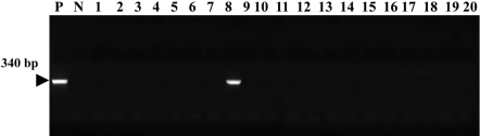
In this study, we examined a colony of 20 beagle dogs in a laboratory animal facility. Mycoplasma was detected by consensus PCR assay in 1 dog with respiratory and constitutional symptoms. None of the other dogs were affected. The dog was euthanized and necropsied. In postmortem examinations, gray or plum-colored gross lesions were found on the lung, most commonly in the apical and cardiac lobes. Some lesions showed clear demarcation and consolidation. Microscopic examination showed peribronchiolar lymphoid hyperplasia and interstitial thickening, lesions pathognomonic for mycoplasma pneumonia. To identify canine Mycoplasma species, we used species-specific PCR reactions for M. arginini, M. canis, M. cynos, M. edwardii, M. felis, M. gateae, M. maculosum, M. molare, M. opalescens, M. spumans, Mycoplasma sp. HRC 689, and M. collis. As the result, we identified Mycoplasma cynos by amplification of DNA extracted from lung tissue of the laboratory beagle dog with respiratory disease.
Keywords: Beagle; Mycoplasma; Mycoplasma cynos; PCR; consensus; dog.
Figures


Similar articles
-
A systematic review and meta-analyses of the association between 4 mycoplasma species and lower respiratory tract disease in dogs.J Vet Intern Med. 2019 Sep;33(5):1880-1891. doi: 10.1111/jvim.15568. Epub 2019 Jul 11. J Vet Intern Med. 2019. PMID: 31297880 Free PMC article.
-
Secreted sialidase activity of canine mycoplasmas.Vet Microbiol. 2009 Jun 12;137(3-4):380-3. doi: 10.1016/j.vetmic.2009.01.009. Epub 2009 Jan 7. Vet Microbiol. 2009. PMID: 19201110 Free PMC article.
-
Canine mycoplasmas: pathogenicity of mycoplasmas associated with distemper pneumonia.J Infect Dis. 1978 Aug;138(2):203-10. doi: 10.1093/infdis/138.2.203. J Infect Dis. 1978. PMID: 681796
-
Detection of Mycoplasma species at various anatomical sites of dogs from different types of kennels.Braz J Microbiol. 2023 Jun;54(2):1251-1255. doi: 10.1007/s42770-023-00947-x. Epub 2023 Mar 18. Braz J Microbiol. 2023. PMID: 36933138 Free PMC article.
-
Canine mycoplasmas.Res Vet Sci. 2005 Aug;79(1):1-8. doi: 10.1016/j.rvsc.2004.10.002. Epub 2004 Nov 24. Res Vet Sci. 2005. PMID: 15894017 Review.
Cited by
-
Development and Validation of a Panel of One-Step Four-Plex qPCR/RT-qPCR Assays for Simultaneous Detection of SARS-CoV-2 and Other Pathogens Associated with Canine Infectious Respiratory Disease Complex.Viruses. 2023 Sep 5;15(9):1881. doi: 10.3390/v15091881. Viruses. 2023. PMID: 37766287 Free PMC article.
-
European surveillance of emerging pathogens associated with canine infectious respiratory disease.Vet Microbiol. 2017 Dec;212:31-38. doi: 10.1016/j.vetmic.2017.10.019. Epub 2017 Oct 28. Vet Microbiol. 2017. PMID: 29173585 Free PMC article.
-
Complete Genome Sequence of Mycoplasma cynos Strain C142.Genome Announc. 2013 Jan;1(1):e00196-12. doi: 10.1128/genomeA.00196-12. Epub 2013 Feb 14. Genome Announc. 2013. PMID: 23409256 Free PMC article.
-
A systematic review and meta-analyses of the association between 4 mycoplasma species and lower respiratory tract disease in dogs.J Vet Intern Med. 2019 Sep;33(5):1880-1891. doi: 10.1111/jvim.15568. Epub 2019 Jul 11. J Vet Intern Med. 2019. PMID: 31297880 Free PMC article.
-
Interstitial pneumonia and pulmonary hypertension associated with suspected ehrlichiosis in a dog.Acta Vet Scand. 2016 Jul 7;58(1):46. doi: 10.1186/s13028-016-0228-1. Acta Vet Scand. 2016. PMID: 27388158 Free PMC article.
References
-
- Fox JG, Anderson LC, Loew FM, Quimby FW. Laboratory Animal Medicine. 2nd ed. Amsterdam: Academic Press; 2002. pp. 111–165.
-
- Rosendal S. Canine mycoplasmas: their ecologic niche and role in disease. J Am Vet Med Assoc. 1982;180(10):1212–1214. - PubMed
LinkOut - more resources
Full Text Sources
Research Materials