. 2012:2012:450738.
doi: 10.1155/2012/450738. Epub 2012 Mar 4.

Adaptive immune response to model antigens is impaired in murine leukocyte-adhesion deficiency-1 revealing elevated activation thresholds in vivo

Affiliations

Adaptive immune response to model antigens is impaired in murine leukocyte-adhesion deficiency-1 revealing elevated activation thresholds in vivo

Thorsten Peters et al. Clin Dev Immunol. 2012.

Abstract

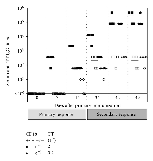
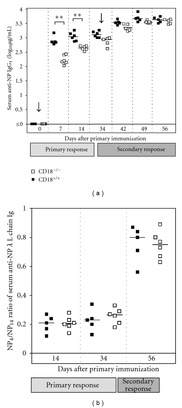
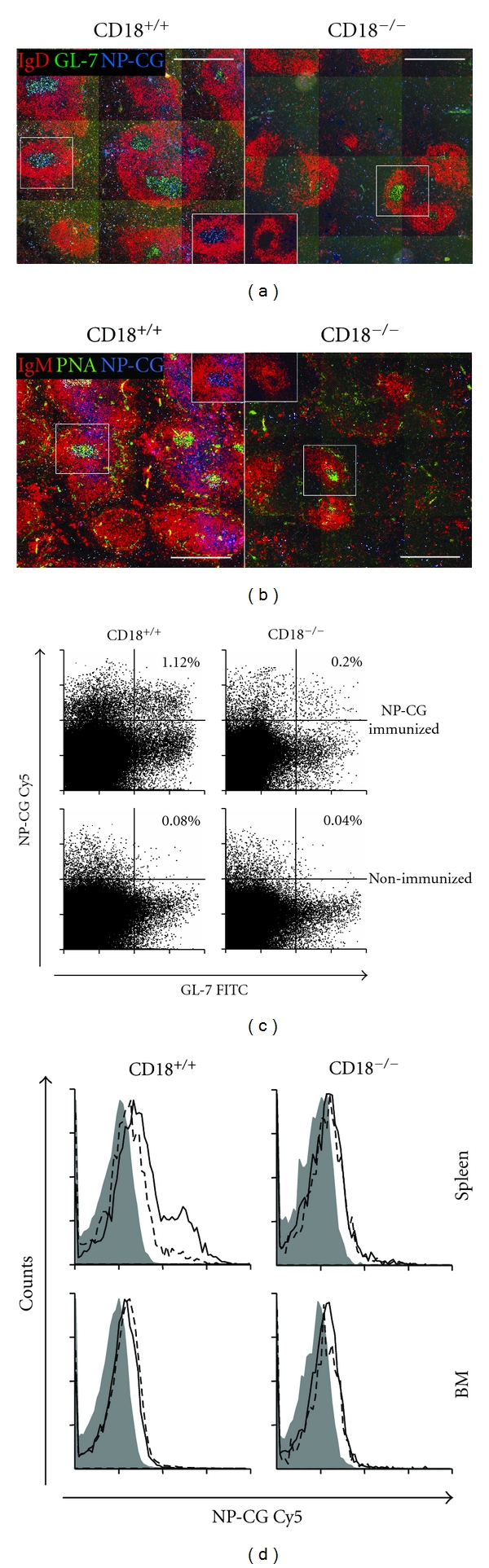
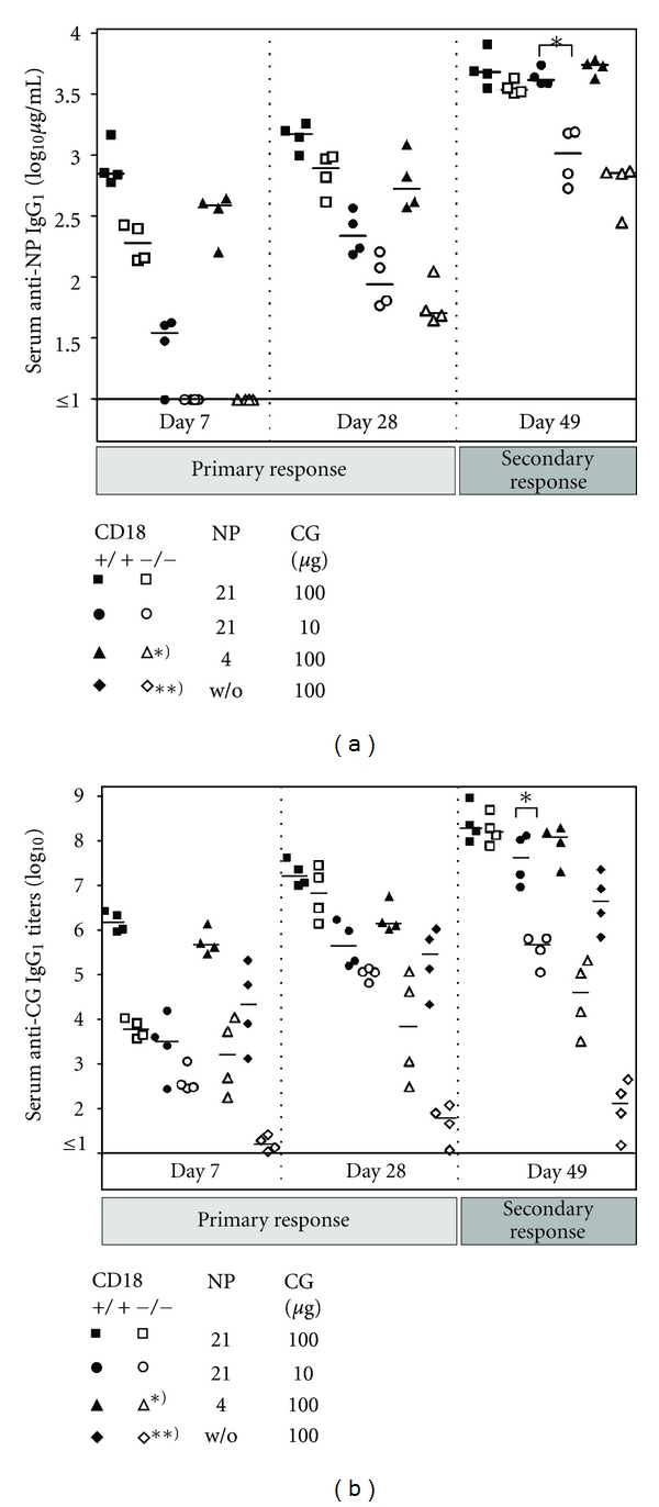
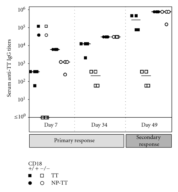
Absence of β₂ integrins (CD11/CD18) leads to leukocyte-adhesion deficiency-1 (LAD1), a rare primary immunodeficiency syndrome. Although extensive in vitro work has established an essential function of β₂ integrins in adhesive and signaling properties for cells of the innate and adaptive immune system, their respective participation in an altered adaptive immunity in LAD1 patients are complex and only partly understood in vivo. Therefore, we investigated adaptive immune responses towards different T-dependent antigens in a murine LAD1 model of β₂ integrin-deficiency (CD18⁻/⁻). CD18⁻/⁻ mice generated only weak IgG responses after immunization with tetanus toxoid (TT). In contrast, robust hapten- and protein-specific immune responses were observed after immunization with highly haptenated antigens such as (4-hydroxy-3-nitrophenyl)₂₁ acetyl chicken γ globulin (NP₂₁-CG), even though regularly structured germinal centers with specificity for the defined antigens/haptens in CD18⁻/⁻ mice remained absent. However, a decrease in the hapten/protein ratio lowered the efficacy of immune responses in CD18⁻/⁻ mice, whereas a mere reduction of the antigen dose was less crucial. Importantly, haptenation of TT with NP (NP-TT) efficiently restored a robust IgG response also to TT. Our findings may stimulate further studies on a modification of vaccination strategies using highly haptenated antigens in individuals suffering from LAD1.


Figures

Figure 1
Figure 1
Defective humoral immune response upon TT in CD18−/− mice. Eight- to twelve-week-old CD18−/− (open symbols) and WT (filled symbols) mice were immunized intraperitoneally with 2.0 (squares) or 0.2 Lf (circles) of tetanus toxoid (TT)/alum. Animals were reimmunized with the same dose of the antigen at day 34. For assessment of the primary immune response, sera were collected at days 0, 7, and 14, for secondary immune response at days 34, 42, and 49. Subsequently, sera were diluted 1 : 10, and plated out on TT-coated plates in 1 : 6 dilution steps. Serum titers of anti-TT specific IgG1 were determined from the last dilution step where the optical density was still above the background level of the assay. Bars represent the median of each group. *Indicates a P < 0.05 for the marked cohorts at all times points shown, from day 14 on.
Figure 2
Figure 2
Functional humoral immune response upon NP-CG in CD18−/− mice. Eight- to twelve-week-old CD18−/− and WT mice were immunized intraperitoneally with 100 μg of alum-precipitated NP-CG. Animals were reinjected with 100 μg of soluble NP-CG at day 34. Sera were collected at days 0, 7, and 14 during primary immune response, and at days 34, 42, 49, and 56 during secondary immune response. (a) Serum levels of anti-NP-specific IgG1 were subsequently detected by ELISA on NP4-coated ELISA plates and calculated by comparison to an IgG1 standard. (b) Anti-NP-specific Ig carrying λ or κ L chains were differentially detected on high-density (NP14-BSA) and low-density (NP4-BSA) hapten-coated ELISA plates in sera obtained at days 14, 34, and 56. Affinity maturation of NP-specific antibodies was estimated as ratio of NP4- to NP14-binding antibodies for each of the three time-points. Bars represent the median of each group. **P < 0.005.
Figure 3
Figure 3
No induction of GC with NP21-CG specificity after immunization in CD18−/− mice. Two weeks after immunization, spleens and BM of WT and CD18−/− mice were subjected to immunofluorescence microscopy (spleens; (a), (b)) or flow cytometry (BM, spleens; (c), (d)). B-cell follicles identify as IgD+ (b) or IgM+ (a) (both in red), whereas GC locate within the follicles and stain IgD GL-7+ (a) or IgM PNA+ (b) (GL-7 and PNA both in green). Specificity for NP-CG was assessed by staining with NP-CG-Cy5 (blue). (a) and (b) show representative sections of spleens solely from immunized mice. Scale bars, 500 μm. Spleen ((c); (d), upper histograms) or BM ((d), lower histograms) cells were analyzed by FACS. (c) displays representative dot plots of splenic cells gated for CD19+ IgMlow. B cells with a GC phenotype additionally stain positive for GL-7 and are situated in the right quadrants of each plot. GC B cells with specificity for NP-CG are located in the upper right quadrants. Percentages for size-gated cells are indicated for immunized (upper dot plots) and mock-immunized (lower dot plots) mice. (d) shows representative histogram plots of cells gated for CD19+ IgMlow GL-7+, representing GC B cells. The continuous lines indicate samples obtained from mice immunized with NP21-CG; dashed lines: mock-immunized mice; grey areas: irrelevant control conjugates.
Figure 4
Figure 4
High NP/CG ratios are critical for elicitation of hapten- and protein-specific IgG titers in CD18−/− mice. Eight- to twelve-week-old CD18−/− (open symbols) and WT (filled symbols) mice were immunized intraperitoneally with 100 μg of NP21-CG/aluM (squares), 10 μg NP21-CG/alum (circles), 100 μg NP4-CG/alum (triangles), or 100 μg CG/alum (diamonds). (a) Serum levels of anti-NP specific IgG1 were subsequently detected by ELISA on NP4-BSA coated ELISA plates and calculated in μg/mL by comparison to IgG1 standards, as described above. (b) For detection of CG-specific IgG1, sera obtained by bleeding from tail veins were diluted 1 : 10, and then plated out on CG-coated plates in 1 : 5 dilution steps. Serum titers of anti-CG-specific IgG1 were determined from the last dilution step where the optical density was still above the background level of the assay. For assessment of the primary immune response, results from sera collected at days 7 and 28 are displayed. Besides, measurements for day 49 are shown, and depict IgG1 titers representative also for further time points assessed during secondary immune responses. Bars represent the median of each group. *P < 0.05; **P < 0.005. Asterisks used in the key box indicate significant differences for the marked cohorts at all times points shown.
Figure 5
Figure 5
Adaptive immunity is functional upon immunization with NP-TT in CD18−/− mice. Eight- to twelve-week-old CD18−/− (open symbols) and WT (filled symbols) mice were immunized intraperitoneally with 2.0 Lf TT/alum (squares) or 2.0 Lf NP-TT/alum. For measurements of anti-TT IgG1, blood was obtained by bleeding from tail veins. Prior to analysis, sera were diluted 1 : 10, and then plated out on TT-coated plates in 1 : 5 dilution steps. Serum titers of anti-TT-specific IgG1 were determined from the last dilution step where the optical density was still above the background level of the assay. For assessment of the primary immune response, results from sera collected at days 7 and 34 are displayed. Besides, measurements for day 49 are shown, and depict IgG1 titers representative also for further time points assessed during secondary immune responses. Bars represent the median of each group.

Similar articles

Cited by

References

    1. Balkow S, Heinz S, Schmidbauer P, et al. LFA-1 activity state on dendritic cells regulates contact duration with T cells and promotes T-cell priming. Blood. 2010;116(11):1885–1894. - PubMed
    1. Carrasco YR, Fleire SJ, Cameron T, Dustin ML, Batista FD. LFA-1/ICAM-1 interaction lowers the threshold of B cell activation by facilitating B cell adhesion and synapse formation. Immunity. 2004;20(5):589–599. - PubMed
    1. Grakoui A, Bromley SK, Sumen C, et al. The immunological synapse: a molecular machine controlling T cell activation. Science. 1999;285(5425):221–227. - PubMed
    1. Iezzi G, Karjalainen K, Lanzavecchia A. The duration of antigenic stimulation determines the fate of naive and effector T cells. Immunity. 1998;8(1):89–95. - PubMed
    1. Perez OD, Mitchell D, Jager GC, et al. Leukocyte functional antigen 1 lowers T cell activation thresholds and signaling through cytohesin-1 and Jun-activating binding protein 1. Nature Immunology. 2003;4(11):1083–1092. - PubMed

Publication types

MeSH terms

LinkOut - more resources