

Antiproliferative and Proapoptotic Effects of Labisia pumila Ethanol Extract and Its Active Fraction in Human Melanoma HM3KO Cells
- PMID: 22474490
- PMCID: PMC3310196
- DOI: 10.1155/2012/123470
Antiproliferative and Proapoptotic Effects of Labisia pumila Ethanol Extract and Its Active Fraction in Human Melanoma HM3KO Cells
Abstract
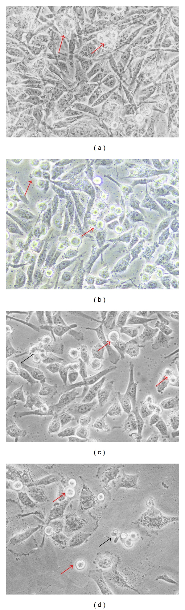
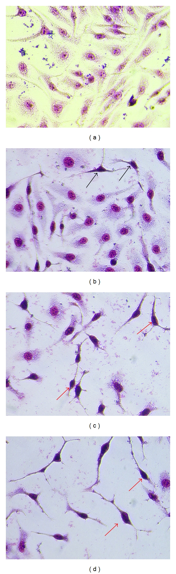
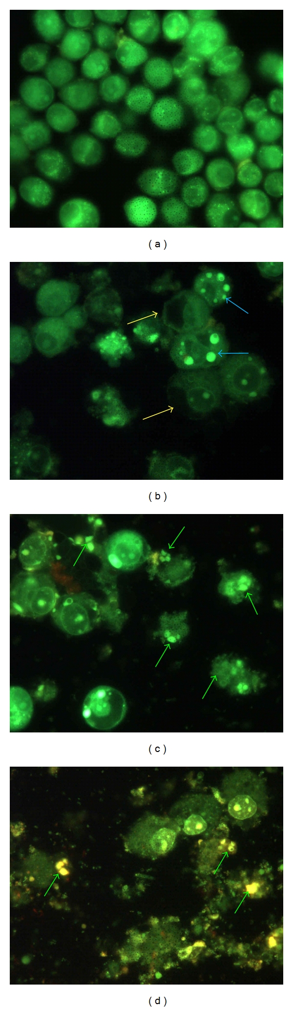
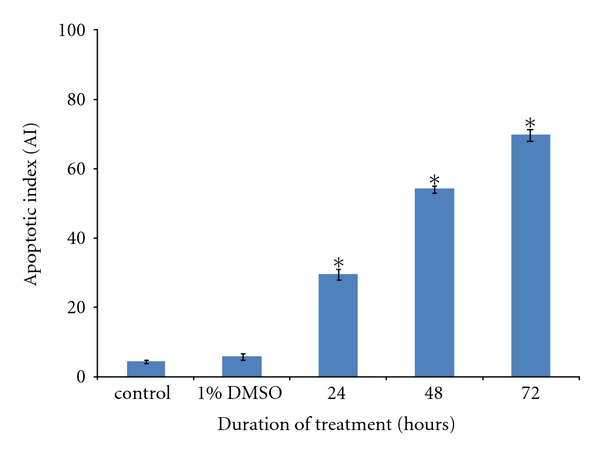
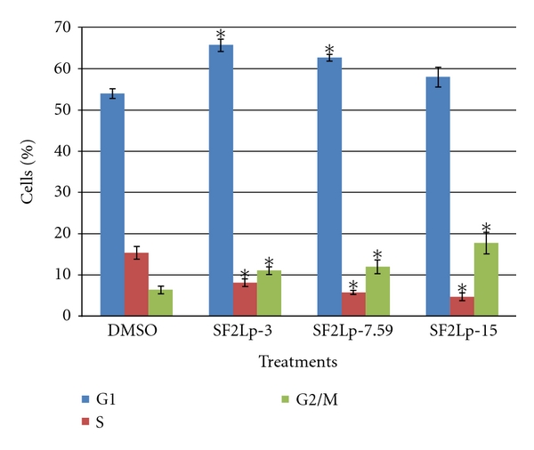
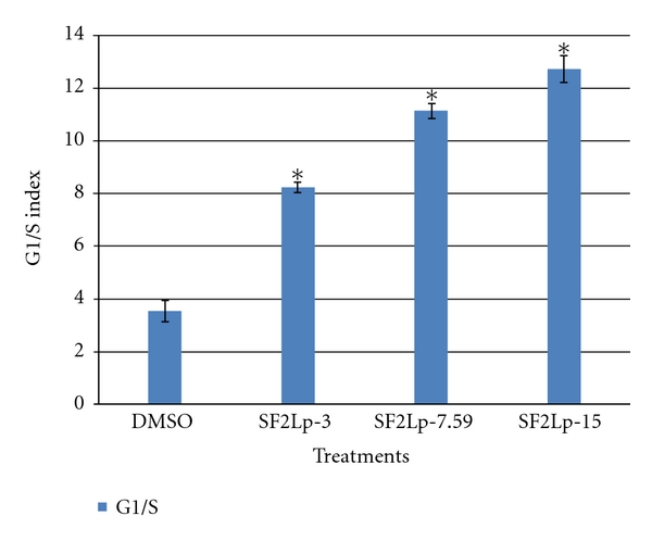
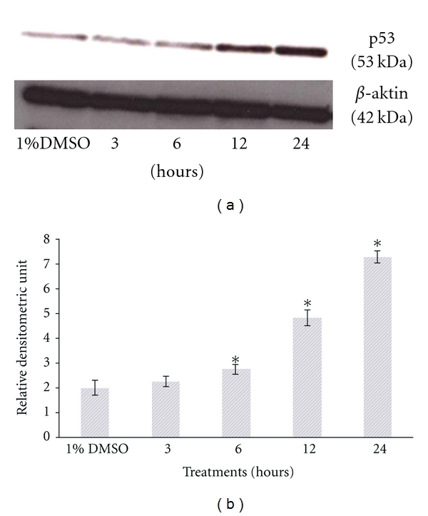
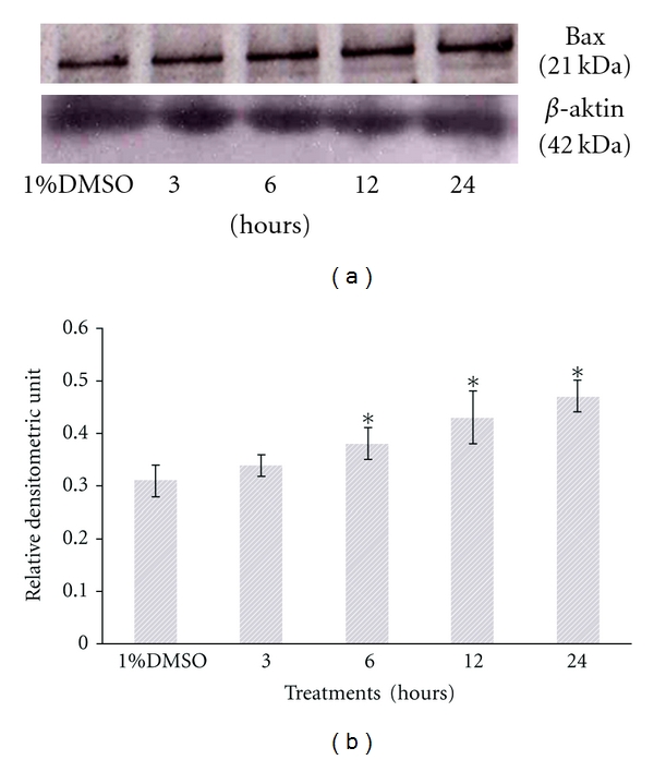
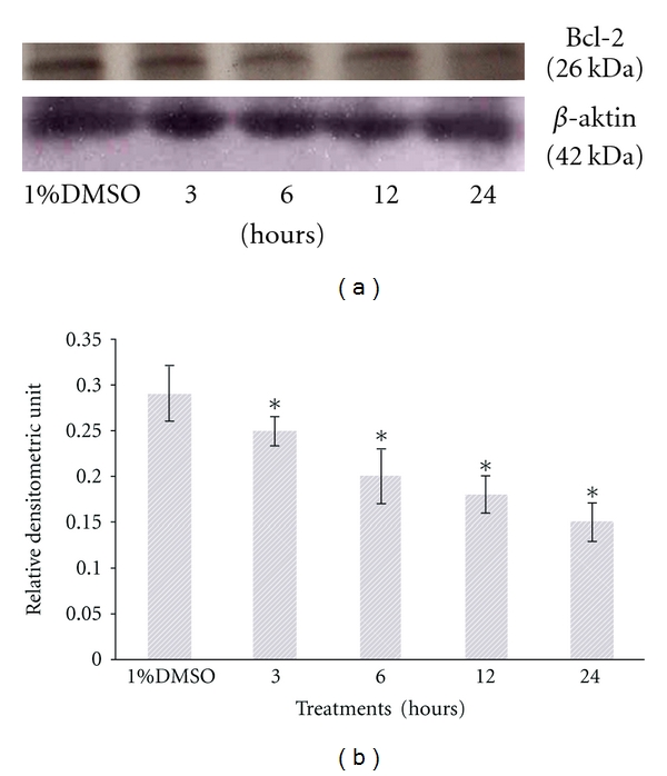
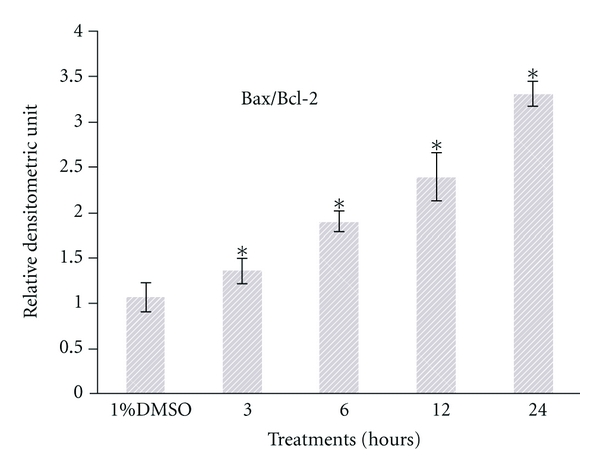
The present study was to determine the anticancer potential of Labisia pumila in in vitro models. Results from the study revealed that ethanol extract of L. pumila was more cytotoxic against HM3KO cells while having reduced effects on nonmalignant cells as compared to aqueous and hexane extracts. Thus, ethanol extract was selected to be further separated by using the bioassay-guided fractionation method to give an active fraction, SF2Lp. Results obtained from the flow cytometry analysis showed that SF2Lp was able to arrest the HM3KO cell cycle at the G1 phase, while morphological findings from AO-EB nuclear staining assays along with the Apoptotic Index confirmed the induction of apoptosis by SF2Lp in HM3KO cells. Results from the mechanistic study further revealed that SF2Lp treatment was able to concurrently increase the expression level of p53 and pro-apoptotic protein Bax and also reduce the expression level of anti-apoptotic protein BCl-2 in HM3KO cells, directly contributing to the increase in Bax/Bcl-2 ratio. These findings, therefore, suggested that L. pumila was able to inhibit HM3KO cell growth possibly by arresting the cell cycle at G1 phase and inducing apoptosis in HM3KO cells via the up- and down-regulation of Bax/Bcl-2 protein, mediated through a p53-dependent pathway.
Figures
Similar articles
-
Anti-Uterine Fibroid Effect of Standardized Labisia Pumila Var. Alata Extracts In Vitro and in Human Uterine Fibroid Cancer Xenograft Model.Asian Pac J Cancer Prev. 2020 Apr 1;21(4):943-951. doi: 10.31557/APJCP.2020.21.4.943. Asian Pac J Cancer Prev. 2020. PMID: 32334454 Free PMC article.
-
In Vitro Pro-Apoptotic and Anti-Migratory Effects of Marantodes pumilum (syn. Labisia pumila) Extracts on Human Prostate Cancer Cell Lines: Bioguided Isolation of 5-Henicosene-1-yl-resorcinol.Plants (Basel). 2023 Apr 6;12(7):1576. doi: 10.3390/plants12071576. Plants (Basel). 2023. PMID: 37050202 Free PMC article.
-
A study of the potential anticancer activity of Mangifera zeylanica bark: Evaluation of cytotoxic and apoptotic effects of the hexane extract and bioassay-guided fractionation to identify phytochemical constituents.Oncol Lett. 2016 Feb;11(2):1335-1344. doi: 10.3892/ol.2016.4087. Epub 2016 Jan 8. Oncol Lett. 2016. PMID: 26893740 Free PMC article.
-
Viscum articulatum Burm. f. aqueous extract exerts antiproliferative effect and induces cell cycle arrest and apoptosis in leukemia cells.J Ethnopharmacol. 2018 Jun 12;219:91-102. doi: 10.1016/j.jep.2018.03.005. Epub 2018 Mar 16. J Ethnopharmacol. 2018. PMID: 29555410
-
Evaluation of anticancer activities of Poria cocos ethanol extract in breast cancer: In vivo and in vitro, identification and mechanism.J Ethnopharmacol. 2020 Jul 15;257:112851. doi: 10.1016/j.jep.2020.112851. Epub 2020 Apr 10. J Ethnopharmacol. 2020. PMID: 32283190
Cited by
-
The Pharmacological Potential of Marantodes pumilum: A Comprehensive Review of Its Medicinal Properties.Int J Mol Sci. 2025 Jun 26;26(13):6155. doi: 10.3390/ijms26136155. Int J Mol Sci. 2025. PMID: 40649931 Free PMC article. Review.
-
Anti-Uterine Fibroid Effect of Standardized Labisia Pumila Var. Alata Extracts In Vitro and in Human Uterine Fibroid Cancer Xenograft Model.Asian Pac J Cancer Prev. 2020 Apr 1;21(4):943-951. doi: 10.31557/APJCP.2020.21.4.943. Asian Pac J Cancer Prev. 2020. PMID: 32334454 Free PMC article.
-
Shoot Multiplication and Callus Induction of Labisia pumila var. alata as Influenced by Different Plant Growth Regulators Treatments and Its Polyphenolic Activities Compared with the Wild Plant.Molecules. 2021 May 27;26(11):3229. doi: 10.3390/molecules26113229. Molecules. 2021. PMID: 34072168 Free PMC article.
-
Influence of cultural practices on breast cancer risks, stage at presentation and outcome in a multi-ethnic developing country.Oncol Lett. 2021 Nov;22(5):806. doi: 10.3892/ol.2021.13067. Epub 2021 Sep 23. Oncol Lett. 2021. PMID: 34630713 Free PMC article. Review.
-
Antiproliferative and apoptosis inducing effect of essential oil extracted from Cyrtomium fortumei (J.) Smith leaves.Med Chem Res. 2015;24(4):1644-1652. doi: 10.1007/s00044-014-1244-1. Epub 2014 Sep 4. Med Chem Res. 2015. PMID: 32214767 Free PMC article.
References
-
- Lee HJ, Lee EO, Rhee YH, et al. An oriental herbal cocktail, ka-mi-kae-kyuk-tang, exerts anti-cancer activities by targeting angiogenesis, apoptosis and metastasis. Carcinogenesis. 2006;27(12):2455–2463. - PubMed
-
- Issa AY, Volate SR, Wargovich MJ. The role of phytochemicals in inhibition of cancer and inflammation: new directions and perspectives. Journal of Food Composition and Analysis. 2006;19(5):405–419.
-
- Cragg GM, Newman DJ. Plants as a source of anti-cancer agents. Journal of Ethnopharmacology. 2005;100(1-2):72–79. - PubMed
-
- Jordan MA, Wilson L. Microtubules as a target for anticancer drugs. Nature Reviews Cancer. 2004;4(4):253–265. - PubMed
-
- Lowe SW, Lin AW. Apoptosis in cancer. Carcinogenesis. 2000;21(3):485–495. - PubMed
LinkOut - more resources
Full Text Sources
Research Materials
Miscellaneous
