. 2012:2012:178178.
doi: 10.1155/2012/178178. Epub 2012 Feb 28.

Activations of Both Extrinsic and Intrinsic Pathways in HCT 116 Human Colorectal Cancer Cells Contribute to Apoptosis through p53-Mediated ATM/Fas Signaling by Emilia sonchifolia Extract, a Folklore Medicinal Plant

Affiliations

Activations of Both Extrinsic and Intrinsic Pathways in HCT 116 Human Colorectal Cancer Cells Contribute to Apoptosis through p53-Mediated ATM/Fas Signaling by Emilia sonchifolia Extract, a Folklore Medicinal Plant

Yu-Hsuan Lan et al. Evid Based Complement Alternat Med. 2012.

Abstract

Emilia sonchifolia (L.) DC (Compositae), an herbaceous plant found in Taiwan and India, is used as folk medicine. The clinical applications include inflammation, rheumatism, cough, cuts fever, dysentery, analgesic, and antibacteria. The activities of Emilia sonchifolia extract (ESE) on colorectal cancer cell death have not been fully investigated. The purpose of this study explored the induction of apoptosis and its molecular mechanisms in ESE-treated HCT 116 human colorectal cancer cells in vitro. The methanolic ESE was characterized, and γ-humulene was formed as the major constituent (63.86%). ESE induced cell growth inhibition in a concentration- and time-dependent response by MTT assay. Apoptotic cells (DNA fragmentation, an apoptotic catachrestic) were found after ESE treatment by TUNEL assay and DNA gel electrophoresis. Alternatively, ESE stimulated the activities of caspase-3, -8, and -9 and their specific caspase inhibitors protected against ESE-induced cytotoxicity. ESE promoted the mitochondria-dependent and death-receptor-associated protein levels. Also, ESE increased ROS production and upregulated the levels of ATM, p53, and Fas in HCT 116 cells. Strikingly, p53 siRNA reversed ESE-reduced viability involved in p53-mediated ATM/Fas signaling in HCT 116 cells. In summary, our result is the first report suggesting that ESE may be potentially efficacious in the treatment of colorectal cancer.


Figures
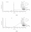
Figure 1
Figure 1
Representative GC-MS analysis of methanolic Emilia sonchifolia extract (ESE) and MS spectra of γ-humulene. The examination conditions and monitoring wavelength of GC-MS analysis were described in the profiles indicated the standard compound (γ-humulene) (a) and ESE (b), respectively.
Figure 2
Figure 2
ESE reduced cell viability and induced morphological changes, apoptosis, and DNA fragmentation in HCT 116 cells. (a) Cells were exposed to various concentrations of ESE (0, 25, 50, 75, or 100 μg/mL) for 24 h and determined and analyzed the viability using the MTT assay. (b) HCT 116 cells in response to 25, 50, and 100 μg/mL of ESE for 24-h exposure were photographed at 200x magnification and showed apoptotic morphological changes. (c) Cells were cultured with 0, 25, 50, 75, or 100 μg/mL of ESE for 24 h for determining apoptotic cells by TUNEL assay and flow cytometric analysis. (d) Treatment with ESE (50 μg/mL) in HCT 116 cells shows DNA ladders by DNA gel electrophoresis. The values presented are the mean ± S.D. (n = 3) from three independent experiments. ***P < 0.001 shows a significant different when compared to control (0 h) sample.
Figure 3
Figure 3
ESE affected the activities and protein levels of caspase-3, -8, and -9 in HCT 116 cells. Cells were treated with 50 μg/mL of ESE for 6, 12, and 24 h. (a) ESE stimulated the activities of caspase-3, caspase-8, and caspase-9 in HCT cells as described in Section 2. The values presented are the mean ± S.D. (n = 3) from three independent experiments. ***P < 0.001 indicates a significant different when compared to control (0 h) sample. (b) The total proteins were harvested from ESE (50 μg/mL) treated HCT 116 cells for 0, 6, 12, and 24 h and determined the protein levels of pro-caspase-3, pro-caspase-8, and pro-caspase-9 by Western blotting. Actin was used as a loading control. (c) ESE stimulated the translocation of caspase-3 trafficking to nuclei in HCT 116 cells by confocal laser scanning microscope as described in Section 2. Cells were pre-incubated with or without specific inhibitors of caspase-3 (Z-DEVD-FMK), caspase-8 (Z-IETD-FMK), and caspase-9 (Z-LEHD-FMK), respectively, and then treated with ESE-(50 μg/mL) for 24 h. The cellular viability was assessed by MTT assay (d) and apoptotic cells were assessed by TUNEL assay and flow cytometric analysis (e). The values presented are the mean ± S.D. (n = 3) from three independent experiments. ***P < 0.001 shows a significant different when compared to ESE treatment.
Figure 4
Figure 4
ESE altered the protein abundance-associated with mitochondria- and death-receptor-dependent apoptotic signaling in HCT 116 cells. Cells were treated with ESE (50 μg/mL) for 0, 6, 12, 24 h, and total protein, cytosolic, and mitochondrial lysates were prepared and subjected to Western blotting analysis. The membranes were incubated with (a) anti-Bcl-2, anti-Bax, anti-Bid and anti-PUMA antibodies; (b) anti-Fas, anti-FasL, anti-DR4 and anti-DR5 antibodies; (c) anticytochrome c antibody. The blot was also probed with anti-Actin and anti-Complex V antibodies to confirm equal loading of samples. Each band was quantified using ImageJ software.
Figure 5
Figure 5
ESE increased ROS production and contributed to p53-correlated ATM/Fas apoptotic signaling in HCT 116 cells. (a) Treatment with ESE (50 μg/mL) for the indicated times (0, 2, and 4 h) was subjected to ROS productions by flow cytometry as described in Section 2. (b) Pretreatment with NAC (10 mM, a scavenger of ROS), or caffeine (1 mM, an inhibitor of ATM) in ESE-treated HCT 116 cells restored the cell viability by MTT assay. The values presented are the mean ± S.D. (n = 3) from three independent experiments. ***P < 0.001 shows a significant different when compared to ESE treatment. (c) ESE elevated the protein levels of ATM, phosphorylated ATM (Ser1981), p53, phosphorylation, and p53 (Ser15) by Western blotting. (d) Effects of ESE on Fas mRNA level in HCT 116 cells, and the total RNA was extracted from each treatment of ESE (50 μg/mL) on HCT 116 cells for 0, 6, and 12 h. RNA samples were reverse transcribed into cDNA and quantified with real-time PCR as described in Section 2. The ratios of Fas mRNA/GAPDH are presented. The values presented are the mean ± S.D. (n = 3) from three independent experiments. ***P < 0.001 shows a significant different when compared to control (0 h) sample.
Figure 6
Figure 6
ESE-affected cytotoxicity and apoptosis is mediated through alterations of p53 downstream signals in p53 siRNA-transfected HCT 116 cells. (a) The p53 siRNA or control siRNA-transfected HCT 116 cells were treated with ESE (50 μg/mL) for 12 h, and total protein was prepared and subjected to Western blotting analysis. The membranes were incubated with anti-p53, anti-Fas, anti-PUMA, anticaspase-8 and, anti-caspase-3 antibodies. The blot was also probed with anti-Actin antibody to confirm equal loading of samples. Each band was quantified using ImageJ software. The p53 siRNA or control siRNA-transfected HCT 116 cells were treated with ESE (50 μg/mL) for 24 h, cell viability was determined by MTT assay (b) and apoptotic cells were assessed by TUNEL assayand flow cytometric analysis (c). The values presented are the mean ± S.D. (n = 3) from three independent experiments. ***P < 0.001 shows a significant different when compared to control sample.
Figure 7
Figure 7
A proposed working model for the action and possible signaling pathways of ESE on HCT 116 human colorectal cancer cells. ESE induces apoptosis through both extrinsic and intrinsic apoptotic pathways, resulting from p53-mediated ATM/Fas signaling, which counteracts the induction of apoptotic death in HCT 116 cells (see text for details).

References

    1. Center MM, Jemal A, Smith RA, Ward E. Worldwide variations in colorectal cancer. CA Cancer Journal for Clinicians. 2009;59(6):366–378. - PubMed
    1. Tan KY, Liu CB, Chen AH, Ding YJ, Jin HY, Seow-Choen F. The role of traditional Chinese medicine in colorectal cancer treatment. Techniques in Coloproctology. 2008;12(1):1–6. - PubMed
    1. Hu B, Shen K-P, An H-M, Wu Y, Du Q. Aqueous extract of curcuma aromatica induces apoptosis and G2/M arrest in human colon carcinoma LS-174-T cells independent of p53. Cancer Biotherapy and Radiopharmaceuticals. 2011;26(1):97–104. - PubMed
    1. Cao B, Li ST, Li Z, Deng WL. Yiqi Zhuyu Decoction Combined with FOLFOX-4 as first-line therapy in metastatic colorectal cancer. Chinese Journal of Integrative Medicine. 2011;17(8):593–599. - PubMed
    1. Cheng WY, Wu SL, Hsiang CY, et al. Relationship between San-Huang-Xie-Xin-Tang and its herbal components on the gene expression profiles in HepG2 cells. American Journal of Chinese Medicine. 2008;36(4):783–797. - PubMed