Review
. 2012 Apr;2(4):a006619.
doi: 10.1101/cshperspect.a006619.

Vascular lumen formation

Affiliations
Review

Vascular lumen formation

Eckhard Lammert et al. Cold Spring Harb Perspect Med. 2012 Apr.

Abstract

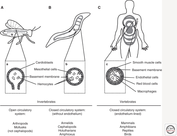
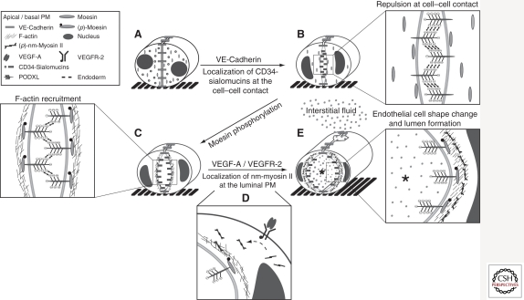
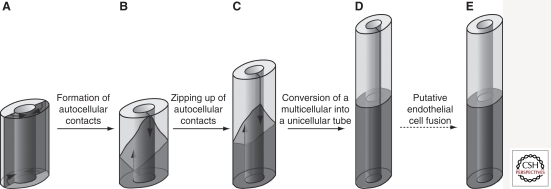
The vascular system developed early in evolution. It is required in large multicellular organisms for the transport of nutrients, oxygen, and waste products to and from tissues. The vascular system is composed of hollow tubes, which have a high level of complexity in vertebrates. Vasculogenesis describes the de novo formation of blood vessels, e.g., aorta formation in vertebrate embryogenesis. In contrast, angiogenesis is the formation of blood vessels from preexisting ones, e.g., sprouting of intersomitic blood vessels from the aorta. Importantly, the lumen of all blood vessels in vertebrates is lined and formed by endothelial cells. In both vasculogenesis and angiogenesis, lumen formation takes place in a cord of endothelial cells. It involves a complex molecular mechanism composed of endothelial cell repulsion at the cell-cell contacts within the endothelial cell cords, junctional rearrangement, and endothelial cell shape change. As the vascular system also participates in the course of many diseases, such as cancer, stroke, and myocardial infarction, it is important to understand and make use of the molecular mechanisms of blood vessel formation to better understand and manipulate the pathomechanisms involved.


Figures

Figure 1.
Figure 1.
Circulatory systems in multicellular organisms. Schematic drawings of representative multicellular organisms. Black dotted lines indicate the place of cross sections shown in the boxes below (a–c). (A) In arthropods and mollusks the organs are surrounded by hemolymph. The heart pumps the hemolymph into the open hemocoel from where it is retrieved back into the heart (arrows). (a) The vascular system is lined with a layer of cardioblasts and a luminal basement membrane. The hemolymph contains hemocytes. (B) In annelids, cephalopods, holothurians, and amphioxus the blood is located in a closed vascular system. The latter consists of blood vessels in which the blood is pumped by contractile myoepithelial cells (arrows show the direction of the blood flow). (b) The vessels are lined by the basement membrane of mesothelial cells and/or intestinal cells. Hemocytes occur free in the vessel lumen as well as adherent at the luminal basement membrane. (C) In vertebrates, the circulatory system is closed. The blood is pumped through the whole organism by the heart. Arrows indicate the direction of blood flow. (c) The vasculature is lined by the apical cell surface of endothelial cells. In contrast, the vascular basement membrane is abluminally located. Peripheral mural cells are located basal, within or at the basement membrane, and stabilize the vessels. The blood mainly contains red blood cells, besides leukocytes and thrombocytes. (Figure is from Strilic et al. 2010; reprinted, with permission, from Springer © 2010.)
Figure 2.
Figure 2.
Molecular mechanism of de novo lumen formation. Schematic drawings of a developing aorta. (A) A part of an endothelial cell cord is shown. ECs are connected via adherens junctions (most prominently vascular endothelial [VE]-cadherin) along the entire endothelial cell–cell contact. The sialomucin PODXL is localized in vesicles. (B) VE-cadherin is involved in localizing negatively charged PODXL to the endothelial cell–cell contact. VE-cadherin itself is translocated to the lateral cell areas. Vesicles with PODXL are released at the cell–cell contact to form an apical cell surface, and the negatively charged sialic acids of PODXL and the other apical glycoproteins lead to a repulsion (arrows) of the apical cell surfaces. This results in the formation of a small slit from which the lumen subsequently develops. (C) After its phosphorylation, moesin and other Ezrin-Radixin-Moesin (ERM) proteins connect PODXL to the F-actin cytoskeleton. (D) VEGF-A activates VEGFR-2, which leads to the phosphorylation of nonmuscular (nm) myosin II and formation of actomyosin complexes. (E) The F-actin cytoskeleton and actomyosin generate the force needed to further separate the apposing apical cell surfaces and thus widen the aortic lumen. (Figure is from Strilic et al. 2009; reprinted, with permission, from Elsevier © 2009.)
Figure 3.
Figure 3.
Transition of multicellular blood vessels into unicellular and seamless blood vessels. (A) A multicellular tube (consisting of a light cell and a dark cell) has a central lumen joined by lateral junctions. The cells reach around the lumen (arrows) to form autocellular cell–cell contacts. (B) When the junctions meet at the opposite sides of the lumen, they start zipping up in two directions (arrows). (C) Autocellular contacts therefore replace the intercellular cell–cell contacts. (D) The conversion of a multicellular tube into a unicellular tube is completed. (E) Conversion of a unicellular into a seamless vascular tube via fusion of the plasma membrane at the autocellular contact sites. (Figure is from Strilic et al. 2010b; reprinted, with permission, from Springer © 2010.)

References

    1. Adachi T, Tomita M, Yoshizato K 2005. Synthesis of prolyl 4-hydroxylase αa subunit and type IV collagen in hemocytic granular cells of silkworm, Bombyx mori: Involvement of type IV collagen in self-defense reaction and metamorphosis. Matrix Biol 24: 136–154 - PubMed
    1. Adams RH, Alitalo K 2007. Molecular regulation of angiogenesis and lymphangiogenesis. Nat Rev Mol Cell Biol 8: 464–478 - PubMed
    1. Barber VC, Graziadei P 1965. The fine structure of cephalopod blood vessels. I. Some smaller peripheral vessels. Z Zellforsch Mikrosk Anat 66: 765–781 - PubMed
    1. Bazzoni G, Dejana E 2004. Endothelial cell-to-cell junctions: Molecular organization and role in vascular homeostasis. Physiol Rev 84: 869–901 - PubMed
    1. Betsholtz C, Lindblom P, Gerhardt H 2005. Role of pericytes in vascular morphogenesis. EXS 115–125 - PubMed

MeSH terms