

Umbilical cord as a mesenchymal stem cell source for treating joint pathologies
- PMID: 22474635
- PMCID: PMC3302041
- DOI: 10.5312/wjo.v2.i6.43
Umbilical cord as a mesenchymal stem cell source for treating joint pathologies
Abstract
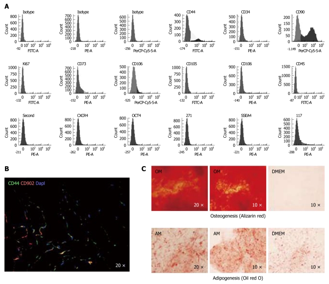
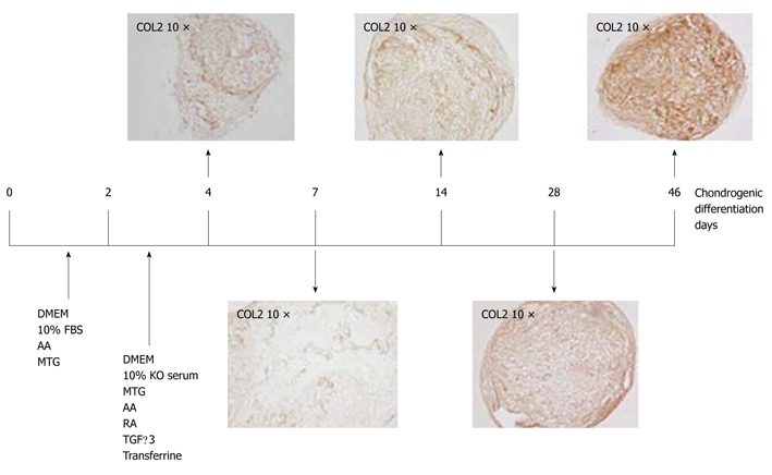
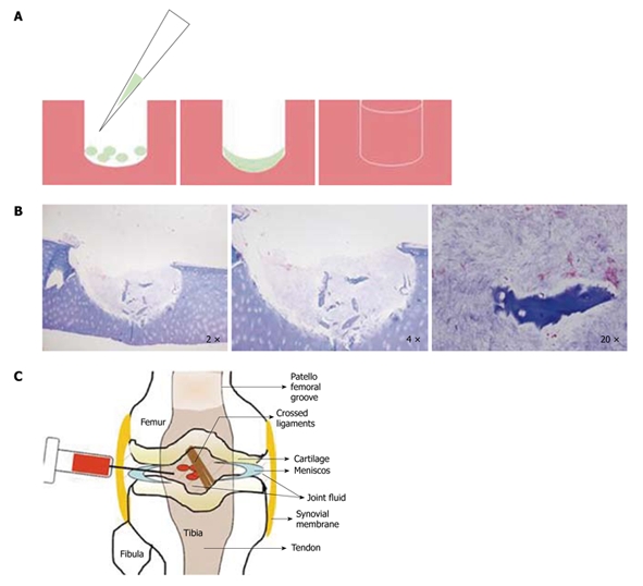
Articular cartilage disorders and injuries often result in life-long chronic pain and compromised quality of life. Regrettably, the regeneration of articular cartilage is a continuing challenge for biomedical research. One of the most promising therapeutic approaches is cell-based tissue engineering, which provides a healthy population of cells to the injured site but requires differentiated chondrocytes from an uninjured site. The use of healthy chondrocytes has been found to have limitations. A promising alternative cell population is mesenchymal stem cells (MSCs), known to possess excellent proliferation potential and proven capability for differentiation into chondrocytes. The "immunosuppressive" property of human MSCs makes them an important candidate for allogeneic cell therapy. The use of allogeneic MSCs to repair large defects may prove to be an alternative to current autologous and allogeneic tissue-grafting procedures. An allogeneic cell-based approach would enable MSCs to be isolated from any donor, expanded and cryopreserved in allogeneic MSC banks, providing a readily available source of progenitors for cell replacement therapy. These possibilities have spawned the current exponential growth in stem cell research in pharmaceutical and biotechnology communities. Our objective in this review is to summarize the knowledge about MSCs from umbilical cord stroma and focus mainly on their applications for joint pathologies.
Keywords: Cartilage degeneration; Human; Mesenchymal stem cell; Umbilical cord.
Figures
Similar articles
-
Mesenchymal stem cells: immunobiology and role in immunomodulation and tissue regeneration.Cytotherapy. 2009;11(4):377-91. doi: 10.1080/14653240903080367. Cytotherapy. 2009. PMID: 19568970 Review.
-
Influence of cellular microenvironment and paracrine signals on chondrogenic differentiation.Front Biosci. 2007 Sep 1;12:4946-56. doi: 10.2741/2440. Front Biosci. 2007. PMID: 17569622 Review.
-
Mesenchymal stem cells in regenerative medicine: Focus on articular cartilage and intervertebral disc regeneration.Methods. 2016 Apr 15;99:69-80. doi: 10.1016/j.ymeth.2015.09.015. Epub 2015 Sep 15. Methods. 2016. PMID: 26384579 Review.
-
Repair of injured articular and growth plate cartilage using mesenchymal stem cells and chondrogenic gene therapy.Curr Stem Cell Res Ther. 2006 May;1(2):213-29. doi: 10.2174/157488806776956904. Curr Stem Cell Res Ther. 2006. PMID: 18220868 Review.
-
Mesenchymal stem cell sheet encapsulated cartilage debris provides great potential for cartilage defects repair in osteoarthritis.Med Hypotheses. 2012 Sep;79(3):420-1. doi: 10.1016/j.mehy.2012.05.024. Epub 2012 Jun 1. Med Hypotheses. 2012. PMID: 22658361
Cited by
-
Implantation of human umbilical cord mesenchymal stem cells for ischemic stroke: perspectives and challenges.Front Med. 2015 Mar;9(1):20-9. doi: 10.1007/s11684-014-0371-x. Epub 2014 Dec 9. Front Med. 2015. PMID: 25491769 Review.
-
Karyotype stability of human umbilical cord-derived mesenchymal stem cells during in vitro culture.Exp Ther Med. 2014 Nov;8(5):1508-1512. doi: 10.3892/etm.2014.1977. Epub 2014 Sep 18. Exp Ther Med. 2014. PMID: 25289050 Free PMC article.
-
Mesenchymal stem cell in vitro labeling by hybrid fluorescent magnetic polymeric particles for application in cell tracking.Med Mol Morphol. 2015 Dec;48(4):204-13. doi: 10.1007/s00795-015-0102-7. Epub 2015 Apr 17. Med Mol Morphol. 2015. PMID: 25893425
-
Mir-218 contributes to the transformation of 5-Aza/GF induced umbilical cord mesenchymal stem cells into hematopoietic cells through the MITF pathway.Mol Biol Rep. 2014 Jul;41(7):4803-16. doi: 10.1007/s11033-014-3351-y. Epub 2014 Apr 3. Mol Biol Rep. 2014. PMID: 24696000
-
Allogeneic Umbilical Cord-Derived Mesenchymal Stem Cells as a Potential Source for Cartilage and Bone Regeneration: An In Vitro Study.Stem Cells Int. 2017;2017:1732094. doi: 10.1155/2017/1732094. Epub 2017 Nov 16. Stem Cells Int. 2017. PMID: 29358953 Free PMC article.
References
-
- Williams JT, Southerland SS, Souza J, Calcutt AF, Cartledge RG. Cells isolated from adult human skeletal muscle capable of differentiating into multiple mesodermal phenotypes. Am Surg. 1999;65:22–26. - PubMed
-
- Prusa AR, Marton E, Rosner M, Bernaschek G, Hengstschläger M. Oct-4-expressing cells in human amniotic fluid: a new source for stem cell research? Hum Reprod. 2003;18:1489–1493. - PubMed
-
- Beyer Nardi N, da Silva Meirelles L. Mesenchymal stem cells: isolation, in vitro expansion and characterization. Handb Exp Pharmacol. 2006:249–282. - PubMed
LinkOut - more resources
Full Text Sources
