

Evaluation of accuracy of 3D reconstruction images using multi-detector CT and cone-beam CT
- PMID: 22474645
- PMCID: PMC3314834
- DOI: 10.5624/isd.2012.42.1.25
Evaluation of accuracy of 3D reconstruction images using multi-detector CT and cone-beam CT
Abstract
Purpose: This study was performed to determine the accuracy of linear measurements on three-dimensional (3D) images using multi-detector computed tomography (MDCT) and cone-beam computed tomography (CBCT).
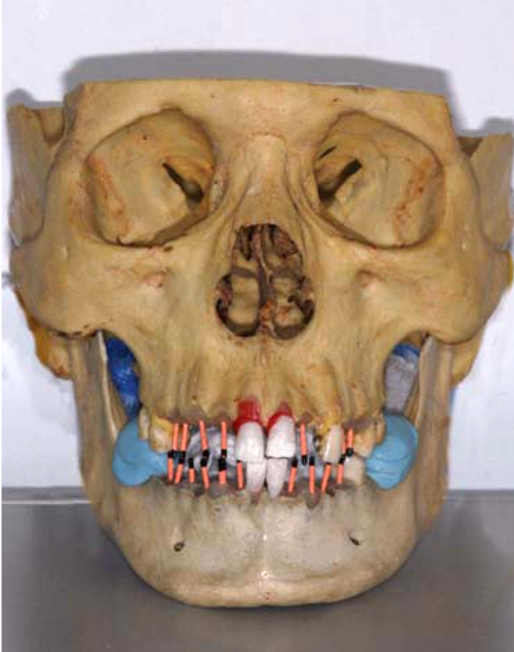
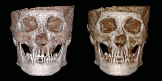
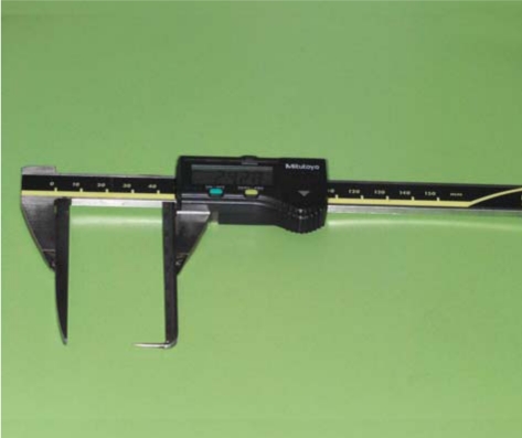
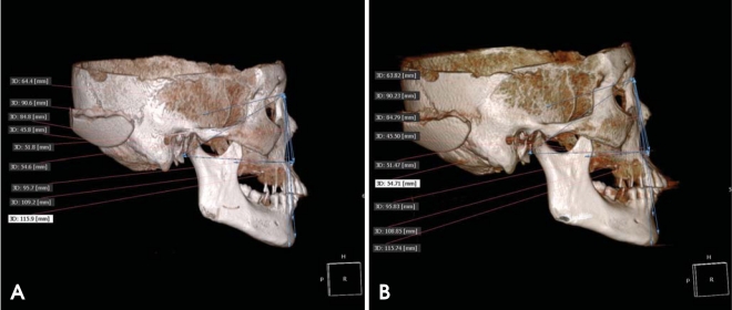
Materials and methods: MDCT and CBCT were performed using 24 dry skulls. Twenty-one measurements were taken on the dry skulls using digital caliper. Both types of CT data were imported into OnDemand software and identification of landmarks on the 3D surface rendering images and calculation of linear measurements were performed. Reproducibility of the measurements was assessed using repeated measures ANOVA and ICC, and the measurements were statistically compared using a Student t-test.
Results: All assessments under the direct measurement and image-based measurements on the 3D CT surface rendering images using MDCT and CBCT showed no statistically difference under the ICC examination. The measurements showed no differences between the direct measurements of dry skull and the image-based measurements on the 3D CT surface rendering images (P>.05).
Conclusion: Three-dimensional reconstructed surface rendering images using MDCT and CBCT would be appropriate for 3D measurements.
Keywords: Reproducibility of Results; Skull; Three-Dimensional Image.
Figures
Similar articles
-
Reliability and accuracy of cone beam computed tomography versus conventional multidetector computed tomography for image-guided craniofacial implant planning: An in vitro study.Int J Oral Maxillofac Implants. 2019 May/June;34(3):665–672. doi: 10.11607/jomi.6915. Epub 2019 Apr 1. Int J Oral Maxillofac Implants. 2019. PMID: 30934042
-
Linear accuracy and reliability of cone beam CT derived 3-dimensional images constructed using an orthodontic volumetric rendering program.Angle Orthod. 2008 May;78(3):387-95. doi: 10.2319/122106-52.1. Angle Orthod. 2008. PMID: 18416632
-
Comparison of linear and angular measurements using two-dimensional conventional methods and three-dimensional cone beam CT images reconstructed from a volumetric rendering program in vivo.Dentomaxillofac Radiol. 2011 Dec;40(8):492-500. doi: 10.1259/dmfr/15644321. Dentomaxillofac Radiol. 2011. PMID: 22065798 Free PMC article.
-
The reliability of cephalometric measurements in oral and maxillofacial imaging: Cone beam computed tomography versus two-dimensional digital cephalograms.Indian J Dent Res. 2016 Jul-Aug;27(4):370-377. doi: 10.4103/0970-9290.191884. Indian J Dent Res. 2016. PMID: 27723632
-
On the data acquisition, image reconstruction, cone beam artifacts, and their suppression in axial MDCT and CBCT - A review.Med Phys. 2018 Jul 17. doi: 10.1002/mp.13095. Online ahead of print. Med Phys. 2018. PMID: 30019342 Review.
Cited by
-
Linear accuracy of cone-beam computed tomography and a 3-dimensional facial scanning system: An anthropomorphic phantom study.Imaging Sci Dent. 2018 Jun;48(2):111-119. doi: 10.5624/isd.2018.48.2.111. Epub 2018 Jun 19. Imaging Sci Dent. 2018. PMID: 29963482 Free PMC article.
-
Three dimensional modeling and quantitative analysis of long bone parameters of rabbit using micro-computed tomography.Iran J Vet Res. 2021 Spring;22(2):140-145. doi: 10.22099/ijvr.2021.39092.5688. Iran J Vet Res. 2021. PMID: 34306112 Free PMC article.
-
Dental 3D Printing Design Based on Neurodegeneration and Virtual Reality Imaging Technology.Biomed Res Int. 2022 Sep 8;2022:6833959. doi: 10.1155/2022/6833959. eCollection 2022. Biomed Res Int. 2022. Retraction in: Biomed Res Int. 2023 Aug 2;2023:9783651. doi: 10.1155/2023/9783651. PMID: 36119937 Free PMC article. Retracted.
-
Sexual dimorphism in human midfacial growth patterns from newborn to 5 years old based on computed tomography.J Anat. 2023 Feb;242(2):132-145. doi: 10.1111/joa.13776. Epub 2022 Oct 8. J Anat. 2023. PMID: 36208113 Free PMC article.
-
Three dimensional (3D) CT reconstruction in cancer imaging.Indian J Med Res. 2013 Jan;137(1):10-1. Indian J Med Res. 2013. PMID: 23481047 Free PMC article. No abstract available.
References
-
- Mozzo P, Procacci C, Tacconi A, Martini PT, Andreis IA. A new volumetric CT machine for dental imaging based on the cone-beam technique: preliminary results. Eur Radiol. 1998;8:1558–1564. - PubMed
-
- Sukovic P. Cone beam computed tomography in craniofacial imaging. Orthod Craniofac Res. 2003;6(suppl 1):31–36. - PubMed
-
- Ludlow JB, Davies-Ludlow LE, Brooks SL, Howerton WB. Dosimetry of 3 CBCT devices for oral and maxillofacial radiology: CB Mercuray, NewTom 3G and i-CAT. Dentomaxillofac Radiol. 2006;35:219–226. - PubMed
-
- Maki K, Inou N, Takanishi A, Miller AJ. Computer-assisted simulations in orthodontic diagnosis and the application of a new cone beam X-ray computed tomography. Orthod Craniofac Res. 2003;6(suppl 1):95–101. - PubMed
-
- Aboudara CA, Hatcher D, Nielsen IL, Miller A. A three-dimensional evaluation of the upper airway in adolescents. Orthod Craniofac Res. 2003;6(suppl 1):173–175. - PubMed
