. 2012 Apr 5;3(4):e292.
doi: 10.1038/cddis.2012.32.

Inhibition of ER stress-mediated apoptosis in macrophages by nuclear-cytoplasmic relocalization of eEF1A by the HIV-1 Nef protein

Affiliations

Inhibition of ER stress-mediated apoptosis in macrophages by nuclear-cytoplasmic relocalization of eEF1A by the HIV-1 Nef protein

W Abbas et al. Cell Death Dis. .

Retraction in

Abstract

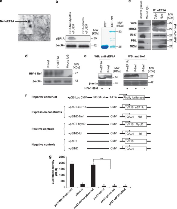
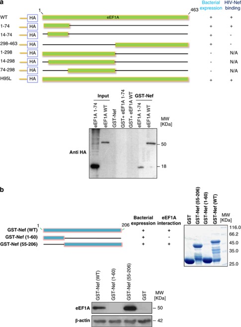
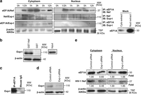
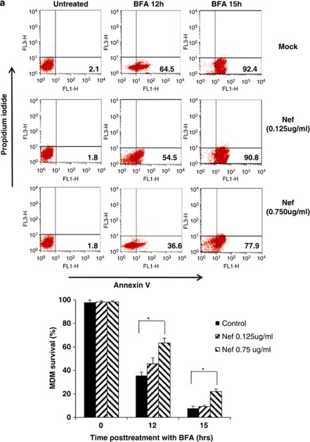
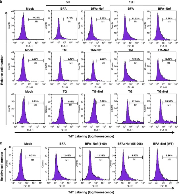
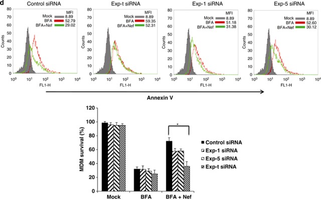
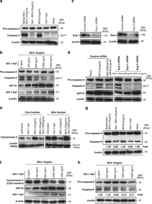
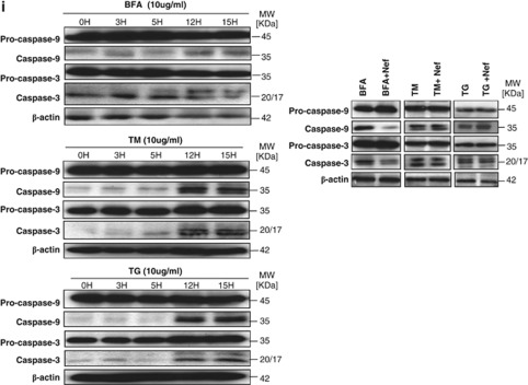
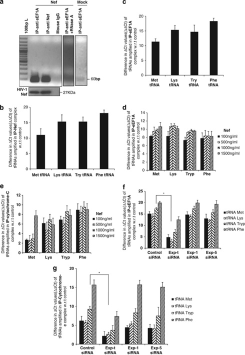
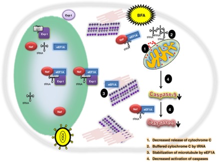
HIV-1 Nef protein has key roles at almost all stages of the viral life cycle. We assessed the role of the Nef/eEF1A (eukaryotic translation elongation factor 1-alpha) complex in nucleocytoplasmic shuttling in primary human macrophages. Nuclear retention experiments and inhibition of the exportin-t (Exp-t) pathway suggested that cytoplasmic relocalization of eEF1A, mediated by Exp-t, occurs in Nef-treated monocyte-derived macrophages (MDMs). We observed the presence of tRNA in the Nef/eEF1A complexes. Nucleocytoplasmic relocalization of the Nef/eEF1A complexes prevented stress-induced apoptosis of MDMs treated with brefeldin-A. Blockade of stress-induced apoptosis of MDMs treated with HIV-1 Nef resulted from enhanced nucleocytoplasmic transport of eEF1A with decreased release of mitochondrial cytochrome c, and from increased tRNA binding to cytochrome c, ultimately leading to an inhibition of caspase activation. Our results indicate that HIV-1 Nef, through the nucleocytoplasmic relocalization of eEF1A and tRNAs, enhances resistance to stress-induced apoptosis in primary human macrophages.


Figures

Figure 1
Figure 1
eEF1A interacts with HIV-1 Nef protein in vitro and in vivo. (a) Image analysis of a high-density protein filter array screened by nonradioactive Far-western blotting using HIV-189.6 Nef as a probe. A magnified section of the blot is shown, indicating the interacting double-spotted eEF1A clones (black). Guide dots (white) on the high-density filter membrane facilitate identification of the xy-coordinates of positive clones. (b) Binding of HIV-1 Nef to eEF1A was measured in GST pull-down assays using U937 cells as a source of lysates. Input corresponds to 10% of the material used for pull-down. β-Actin detection represents input loading controls of the lystates that were used in binding reactions. Results are representative of three independent experiments. The right panel represents Commassie staining of expressed proteins that were used in the binding reaction of GST pull-down. (c) Cytoplasmic and nuclear extracts from several cell lines (Vero, MRC5, U937 cells), PBLs, and MDMs treated with rNef (100 ng/ml) for 0.5 h were immunoprecipitated with an anti-eEF1A antibody, or Nef (100 ng/ml)-treated total cellular lysates were immunoprecipitated with an isotype control antibody. Immunoprecipitated material was analyzed by western blotting with an anti-Nef monoclonal antibody. Recombinant Nef-treated total cell lysates were used as a positive control. Results are representative of three independent experiments. (d) Cellular extracts of MDMs transfected for 48 h with a nef-expressing plasmid (pNef) were immunoprecipitated with an anti-eEF1A antibody, anti-Nef antibody, or isotype control antibody. Immunoprecipitated material was analyzed by western blotting with an anti-Nef monoclonal antibody. Results are representative of two independent experiments. (e) Cellular extracts of PBMCs infected in vitro with HIV-189.6 or mock infected were immunoprecipitated with an anti-eEF1A antibody or anti-Nef monoclonal antibody. Immunoprecipitated material was analyzed by western blotting with an anti-Nef monoclonal antibody or anti-eEF1A antibody. Results are representative of two independent experiments. (f and g) eEF1A and HIV-1 Nef interact in vitro in a mammalian two-hybrid assay. (f) Schematic representation of expression constructs used in co-transfection experiments in the mammalian two-hybrid model. (g) Mammalian two-hybrid analysis with eEF1A fused to the VP16 activator domain and HIV-1 Nef fused to the GAL4 domain. Luciferase assays were conducted on total extracts from U937 cells transfected with the luciferase expression construct pG5–Luc, pBIND–Nef, pACT–eEF1A or control plasmids. Results represent the average of a triplicate experiment in which luciferase was normalized to protein expression. As a positive control, two plasmids, pACT-MyoD and pBIND-Id, were co-transfected, and co-transfection of empty vectors was used as a negative control. Results represent the mean of three independent experiments. *** P<0.001
Figure 2
Figure 2
The N-terminal 74 amino acids of eEF1A binds to the C-terminal region (aa 55–206) of HIV-1 Nef. (a) The N-terminal region is sufficient for binding to HIV-1 Nef. Upper panel, Schematic diagram of eEF1A mutants expressed in bacteria as HA-tagged fusion proteins. The names of the mutants are shown to the left of the figure; numbers refer to the amino acid residues retained by the deletion mutants. The bacterial expression status and in vitro binding of these mutants are reported qualitatively as +or − to the right of the figure. NA, not applicable; WT, wild type. Lower panel, eEF1A interacts with HIV-1 Nef via its N terminus extremity (aa 1–74). Using wild-type GST–Nef constructs, binding of purified WT eEF1A and eEF1A 1–74 was measured in GST pull-down assays. Input corresponds to 10% of the material used for pull-down. Results are representative of two independent experiments. (b) The C-terminal region (aa 55–206) of HIV-1 Nef is sufficient for binding to eEF1A. Upper left panel, Schematic diagram of WT HIV-1 Nef and mutants expressed in bacteria as GST-tagged fusion proteins. The names of the mutants are shown to the left of the figure; numbers refer to the amino-acid residues retained by the deletion mutants. The bacterial expression status and in vitro binding of these mutants are reported qualitatively as + or − to the right of the figure. Upper right panel represents Commassie staining of expressed proteins that were used in the binding reaction of GST pull-down. Lower panel, HIV-1 Nef interacts with eEF1A via its C-terminal region (aa 55–206). The binding of eEF1A present in lysates of MDMs was measured in GST pull-down assays using WT and mutated GST–Nef constructs. Input corresponds to 10% of the material used for pull-down. Results are representative of two independent experiments
Figure 3
Figure 3
Nuclear-cytoplasmic relocalization of eEF1A/rNef complexes occurs in MDMs treated with rNef and depends on Exp-t. (a) Kinetics of eEF1A/rNef/Exp-t complexes in nuclear and cytoplasmic compartments of MDMs. Nuclear and cytoplasmic extracts of MDMs treated with rNef (100 ng/ml) were prepared and the eEF1A/rNef, rNef/Exp-t and eEF1A/Exp-t interactions assessed in both cellular compartments up to 12 h post-treatment using co-immunoprecipitation and western blotting. Similarly, nuclear and cytoplasmic extracts of untreated MDMs (Mock) were prepared and tested for the expression of eEF-1A. β-Actin and TBP were used as loading controls. Results are representative of three independent experiments. (b) Using wild-type GST–Nef constructs, the binding of Exp-t present in MDM lysates was assessed in GST pull-down assays. Input corresponds to 10% of the material used for pull-down. Results are representative of two independent experiments. (c) eEF1A interacts with Exp-t in MDM lysates. Total MDM extracts were prepared and the eEF1A/Exp-t interaction assessed by immunoprecipitation with an anti-eEF1A antibody and western blotting with an anti-Exp-t monoclonal antibody. Results are representative of three independent experiments. (d) Knockdown of Exp-t protein by siRNA in MDMs. MDM cultures were transfected with scrambled control or Exp-t siRNA and total cellular extracts prepared 48 h post-transfection. Protein expression was analyzed by western blot. β-Actin was used as a loading control. (e) Effects of Exp-t siRNA on nuclear-cytoplasmic transport of eEF1A and rNef in MDMs. MDM cultures were transfected with a scrambled control or Exp-t siRNA for 48 h before treatment with rNef (100 ng/ml) for 3 h. Nuclear and cytoplasmic extracts were prepared and analyzed by western blot using anti-Nef and anti-eEF1A antibodies. β-Actin and TBP were used as loading controls. Results are representative of two independent experiments. Protein levels of eEF1A and Nef after siRNA transfection were quantified by densitometry using ImageJ 1.40 software (protein levels in cells transfected with scrambled siRNAs were arbitrarily established at 1)
Figure 4
Figure 4
rNef-mediated inhibition of BFA-induced apoptosis in MDMs parallels cytoplasmic accumulation of eEF1A and is dependent on Exp-t. (a) rNef prevents BFA-induced apoptosis in MDMs. MDMs were treated with BFA (10 μg/ml) for 12 or 15 h or untreated in the presence of increasing concentrations of rNef (0, 125, 750 ng/ml). Apoptosis was detected by flow cytometric analysis of annexin-V. The histogram summarizes the survival of MDMs following treatment with BFA (10 μg/ml) for 12 or 15 h in the presence of increasing concentrations of rNef. The results represent means of three independent experiments; *P<0.05. (b) rNef prevents BFA-induced apoptosis, but neither TM-induced apoptosis nor TG-induced apoptosis in MDMs. MDMs were treated with BFA (10 μg/ml), TM (10 μg/ml), or TG (10 μg/ml) for 5 h or 12 h or untreated in the presence of rNef (1 μg/ml). Apoptosis was measured by TUNEL assay. Results are representative of data observed in three independent experiments. (c) The C-terminal extremity of Nef prevents BFA-induced apoptosis in MDMs. MDMs were treated with BFA (10 μg/ml) for 12 h or untreated in the presence of WTNef, Nef1-60, or Nef55-206 (1 μg/ml). Apoptosis was measured by TUNEL assay. Results are representative of data observed in two independent experiments. (d) Blockade of BFA-induced apoptosis in MDMs by rNef is dependent on Exp-t. MDM cultures were transfected with a scrambled control, Exp-t siRNA, Exp-1 siRNA or Exp-5 siRNA for 48 h before treatment with BFA (10 μg/ml) for 12 h or untreated, in the presence (1 μg/ml) or absence of rNef. Apoptosis was detected by flow cytometric analysis of annexin-V. The histogram shows the survival percentage of MDMs treated with mock, BFA or BFA+Nef in the presence of exportin siRNAs. Results are representative of three independent experiments; *P<0.05
Figure 4
Figure 4
rNef-mediated inhibition of BFA-induced apoptosis in MDMs parallels cytoplasmic accumulation of eEF1A and is dependent on Exp-t. (a) rNef prevents BFA-induced apoptosis in MDMs. MDMs were treated with BFA (10 μg/ml) for 12 or 15 h or untreated in the presence of increasing concentrations of rNef (0, 125, 750 ng/ml). Apoptosis was detected by flow cytometric analysis of annexin-V. The histogram summarizes the survival of MDMs following treatment with BFA (10 μg/ml) for 12 or 15 h in the presence of increasing concentrations of rNef. The results represent means of three independent experiments; *P<0.05. (b) rNef prevents BFA-induced apoptosis, but neither TM-induced apoptosis nor TG-induced apoptosis in MDMs. MDMs were treated with BFA (10 μg/ml), TM (10 μg/ml), or TG (10 μg/ml) for 5 h or 12 h or untreated in the presence of rNef (1 μg/ml). Apoptosis was measured by TUNEL assay. Results are representative of data observed in three independent experiments. (c) The C-terminal extremity of Nef prevents BFA-induced apoptosis in MDMs. MDMs were treated with BFA (10 μg/ml) for 12 h or untreated in the presence of WTNef, Nef1-60, or Nef55-206 (1 μg/ml). Apoptosis was measured by TUNEL assay. Results are representative of data observed in two independent experiments. (d) Blockade of BFA-induced apoptosis in MDMs by rNef is dependent on Exp-t. MDM cultures were transfected with a scrambled control, Exp-t siRNA, Exp-1 siRNA or Exp-5 siRNA for 48 h before treatment with BFA (10 μg/ml) for 12 h or untreated, in the presence (1 μg/ml) or absence of rNef. Apoptosis was detected by flow cytometric analysis of annexin-V. The histogram shows the survival percentage of MDMs treated with mock, BFA or BFA+Nef in the presence of exportin siRNAs. Results are representative of three independent experiments; *P<0.05
Figure 5
Figure 5
rNef-mediated cytoplasmic accumulation of eEF1A in BFA-treated MDMs inhibits caspase activation and decreases the cytoplasmic release of cytochrome c. (a) Inhibition of caspase-3 activation in BFA-stimulated MDMs treated with rNef (1000 ng/ml). (b) Inhibition of caspase-3 activation in BFA-stimulated MDMs treated with rNef is dose-dependent and positively correlates with cytoplasmic accumulation of eEF1A. (c) Knockdown of Exp-1 and Exp-5 proteins by siRNA in MDMs. MDM cultures were transfected with scrambled control, Exp-1 siRNA or Exp-5 siRNA. Total cellular extracts were prepared 48 h post-transfection. Protein expression was analyzed by western blot. β-Actin was used as a loading control. (d) Inhibition of caspase-3 activation in BFA-stimulated MDMs treated with rNef is dependent on Exp-t. (e) Mitochondrial cytochrome c release in BFA-treated MDMs is blocked by rNef. (f) Mitochondrial cytochrome c release in BFA-treated MDMs is blocked by rNef in a dose-dependent manner and positively correlates with cytoplasmic accumulation of eEF1A. (g) Inhibition of caspase-9 activation in BFA-stimulated MDMs treated with rNef (1000 ng/ml). Protein levels of caspase-9 were quantified by densitometry using ImageJ 1.40 software (the level of caspase-9 in mock cells was arbitrarily established at 1). (h) Inhibition of caspase-9 activation in BFA-stimulated MDMs treated with rNef is dose-dependent. Protein levels of caspase-9 were quantified by densitometry using ImageJ 1.40 software (the level of caspase-9 in mock cells was arbitrarily established at 1). (i) Inhibition of caspase activation in BFA-stimulated MDMs treated with rNef, but neither in TM-stimulated MDMs nor in TG-stimulated MDMs treated with rNef. Left panel, Time course of caspase activation in BFA-stimulated MDMs, TM-stimulated MDMs, or TG-stimulated MDMs. Right panel, Inhibition of caspase activation in BFA-stimulated MDMs treated with rNef, but neither in TM-stimulated MDMs nor in TG-stimulated MDMs treated with rNef. MDM cultures were treated with BFA (10 μg/ml), TM (10 μg/ml) or TG (10 μg/ml) for 12h in the presence of rNef (1 μg/ml), and caspase-3 and caspase-9 activation was measured in total cellular lysates. Results are representative of data obtained in three independent experiments
Figure 5
Figure 5
rNef-mediated cytoplasmic accumulation of eEF1A in BFA-treated MDMs inhibits caspase activation and decreases the cytoplasmic release of cytochrome c. (a) Inhibition of caspase-3 activation in BFA-stimulated MDMs treated with rNef (1000 ng/ml). (b) Inhibition of caspase-3 activation in BFA-stimulated MDMs treated with rNef is dose-dependent and positively correlates with cytoplasmic accumulation of eEF1A. (c) Knockdown of Exp-1 and Exp-5 proteins by siRNA in MDMs. MDM cultures were transfected with scrambled control, Exp-1 siRNA or Exp-5 siRNA. Total cellular extracts were prepared 48 h post-transfection. Protein expression was analyzed by western blot. β-Actin was used as a loading control. (d) Inhibition of caspase-3 activation in BFA-stimulated MDMs treated with rNef is dependent on Exp-t. (e) Mitochondrial cytochrome c release in BFA-treated MDMs is blocked by rNef. (f) Mitochondrial cytochrome c release in BFA-treated MDMs is blocked by rNef in a dose-dependent manner and positively correlates with cytoplasmic accumulation of eEF1A. (g) Inhibition of caspase-9 activation in BFA-stimulated MDMs treated with rNef (1000 ng/ml). Protein levels of caspase-9 were quantified by densitometry using ImageJ 1.40 software (the level of caspase-9 in mock cells was arbitrarily established at 1). (h) Inhibition of caspase-9 activation in BFA-stimulated MDMs treated with rNef is dose-dependent. Protein levels of caspase-9 were quantified by densitometry using ImageJ 1.40 software (the level of caspase-9 in mock cells was arbitrarily established at 1). (i) Inhibition of caspase activation in BFA-stimulated MDMs treated with rNef, but neither in TM-stimulated MDMs nor in TG-stimulated MDMs treated with rNef. Left panel, Time course of caspase activation in BFA-stimulated MDMs, TM-stimulated MDMs, or TG-stimulated MDMs. Right panel, Inhibition of caspase activation in BFA-stimulated MDMs treated with rNef, but neither in TM-stimulated MDMs nor in TG-stimulated MDMs treated with rNef. MDM cultures were treated with BFA (10 μg/ml), TM (10 μg/ml) or TG (10 μg/ml) for 12h in the presence of rNef (1 μg/ml), and caspase-3 and caspase-9 activation was measured in total cellular lysates. Results are representative of data obtained in three independent experiments
Figure 6
Figure 6
rNef-mediated cytoplasmic accumulation of eEF1A in BFA-treated MDMs parallels tRNA binding to cytochrome c. (a) Direct detection of tRNAs in rNef/eEF1A complexes in MDMs treated with rNef. Lysates from MDMs treated with rNef (100 ng/ml) for 3 h or left untreated (Mock) were prepared, immunoprecipitated with anti-eEF1A and anti-Nef antibodies, and tRNALys in the eEF1A/rNef complexes amplified by RT-PCR. Lysates were also immunoprecipitated with isotype control antibody. The presence of HIV-1 Nef protein in the immune complex was determined by western blot. As an internal control, lysates from MDMs treated with rNef (100 ng/ml) for 3 h were prepared, treated with RNAse A (10 μg/ml) for 30 min, immunoprecipitated with an anti-eEF1A antibody, and tRNALys in the eEF1A/Nef complexes was amplified by RT-PCR. (b and c) MDMs were treated with rNef (100 ng/ml); the lysates were immunoprecipitated with an anti-Nef Ab (b) or anti-eEF1A Ab (c) and the presence of tRNAMet, tRNALys, tRNATry and tRNAPhe in the Nef/eEF1A complex determined by qRT-PCR. (d and e) MDMs were treated with BFA for 12 h in the presence or absence of rNef (0–1500 ng/ml); the lysates were immunoprecipitated with an anti-eEF1A Ab (d) or anti-cytochrome c Ab (e), and the presence of tRNAMet, tRNALys, tRNATry, and tRNAPhe was determined by qRT-PCR. (f and g) The presence of tRNAs associated with eEF1A and cytochrome c in BFA-stimulated MDMs treated with rNef is dependent on Exp-t. MDM cultures were transfected with scrambled control siRNA, Exp-t siRNA, Exp-1 siRNA or Exp-5 siRNA. Cytoplasmic extracts of MDMs treated with BFA (10 μg/ml) for 12 h in the absence or presence of rNef (1000 ng/ml) were prepared 48 h post-transfection, immunoprecipitated with an anti-eEF1A Ab (f) or anti-cytochrome c Ab (g), and the presence of tRNALys, tRNAMet, tRNAPhe, and tRNATry binding to eEF1A and cytochrome c was determined using qRT-PCR. Results representative of three independent experiments are shown; * P< 0.05
Figure 7
Figure 7
Potential inhibitory effect of the eEF1A/rNef/tRNA complex on the intrinsic apoptotic pathway in BFA-treated MDMs. HIV-1 Nef protein favors translocation and accumulation of eEF1A from nucleus toward to the cytoplasm of the cell. tRNA is present in the Nef/eEF1A complex, which is exported by exportin-t and buffers the cytochrome c released under oxidative stress conditions. At the same time the Nef/eEF1A complex inhibits the caspase activation. Further, eEF1A could stabilize the microtubules and give relief to the cell to survive under stress conditions. BFA, brefeldin-A; Expt, exportin-t

Similar articles

Cited by

References

    1. Walter P, Ron D. The unfolded protein response: from stress pathway to homeostatic regulation. Science. 2011;334:1081–1086. - PubMed
    1. Shore GC, Papa FR, Oakes SA. Signaling cell death from the endoplasmic reticulum stress response. Curr Opin Cell Biol. 2011;23:143–149. - PMC - PubMed
    1. Elbim C, Pillet S, Prevost MH, Preira A, Girard PM, Rogine N, et al. Redox and activation status of monocytes from human immunodeficiency virus-infected patients: relationship with viral load. J Virol. 1999;73:4561–4566. - PMC - PubMed
    1. Masutani H, Ueda S, Yodoi J. The thioredoxin system in retroviral infection and apoptosis. Cell Death Diff. 2005;12:991–998. - PubMed
    1. Uhm HD, Orenstein JM, Wahl SM. Fas mediates apoptosis in human monocytes by a reactive oxygen intermediate dependent pathway. J Immunol. 1996;156:3469–3477. - PubMed

Publication types

MeSH terms