

Paraspeckle nuclear bodies--useful uselessness?
- PMID: 22476590
- PMCID: PMC3428521
- DOI: 10.1007/s00018-012-0973-x
Paraspeckle nuclear bodies--useful uselessness?
Abstract
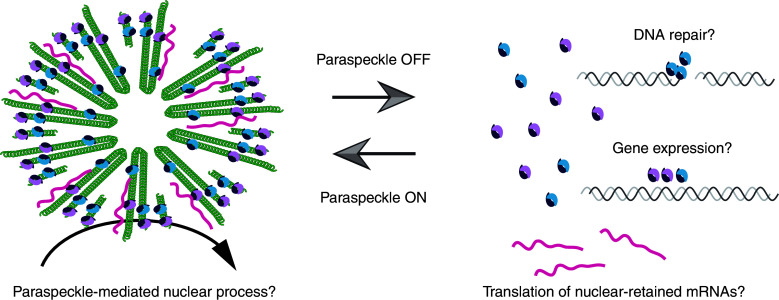
The nucleus of higher eukaryotes, such as humans and mice, is compartmentalized into multiple nuclear bodies, an organization that allows for the regulation of complex gene expression pathways that are characteristic of these organisms. Paraspeckles are recently discovered, mammalian-specific nuclear bodies built on a long, non-protein-coding RNA, NEAT1 (nuclear-enriched abundant transcript 1), which assembles various protein components including RNA-binding proteins of the DBHS (Drosophila behavior and human splicing) family. Paraspeckles have been proposed to control several biological processes, such as stress responses and cellular differentiation, but their function at the whole animal level remains unclear. In this review, we summarize a series of studies on paraspeckles that have been carried out in the decade since their discovery and discuss their physiological function and molecular mechanism.
Figures
Similar articles
-
Paraspeckles: nuclear bodies built on long noncoding RNA.J Cell Biol. 2009 Sep 7;186(5):637-44. doi: 10.1083/jcb.200906113. Epub 2009 Aug 31. J Cell Biol. 2009. PMID: 19720872 Free PMC article. Review.
-
Direct visualization of the co-transcriptional assembly of a nuclear body by noncoding RNAs.Nat Cell Biol. 2011 Jan;13(1):95-101. doi: 10.1038/ncb2140. Epub 2010 Dec 19. Nat Cell Biol. 2011. PMID: 21170033 Free PMC article.
-
An architectural role for a nuclear noncoding RNA: NEAT1 RNA is essential for the structure of paraspeckles.Mol Cell. 2009 Mar 27;33(6):717-26. doi: 10.1016/j.molcel.2009.01.026. Epub 2009 Feb 12. Mol Cell. 2009. PMID: 19217333 Free PMC article.
-
The long noncoding RNA Neat1 is required for mammary gland development and lactation.RNA. 2014 Dec;20(12):1844-9. doi: 10.1261/rna.047332.114. Epub 2014 Oct 14. RNA. 2014. PMID: 25316907 Free PMC article.
-
Organization and function of paraspeckles.Essays Biochem. 2020 Dec 7;64(6):875-882. doi: 10.1042/EBC20200010. Essays Biochem. 2020. PMID: 32830222 Review.
Cited by
-
mRNP granules. Assembly, function, and connections with disease.RNA Biol. 2014;11(8):1019-30. doi: 10.4161/15476286.2014.972208. RNA Biol. 2014. PMID: 25531407 Free PMC article. Review.
-
The DBHS proteins SFPQ, NONO and PSPC1: a multipurpose molecular scaffold.Nucleic Acids Res. 2016 May 19;44(9):3989-4004. doi: 10.1093/nar/gkw271. Epub 2016 Apr 15. Nucleic Acids Res. 2016. PMID: 27084935 Free PMC article.
-
Dynamic integration of splicing within gene regulatory pathways.Cell. 2013 Mar 14;152(6):1252-69. doi: 10.1016/j.cell.2013.02.034. Cell. 2013. PMID: 23498935 Free PMC article. Review.
-
Nono deficiency compromises TET1 chromatin association and impedes neuronal differentiation of mouse embryonic stem cells.Nucleic Acids Res. 2020 May 21;48(9):4827-4838. doi: 10.1093/nar/gkaa213. Nucleic Acids Res. 2020. PMID: 32286661 Free PMC article.
-
Circadian RNA expression elicited by 3'-UTR IRAlu-paraspeckle associated elements.Elife. 2016 Jul 21;5:e14837. doi: 10.7554/eLife.14837. Elife. 2016. PMID: 27441387 Free PMC article.
References
-
- Platani M, Lamond AI (2008) Nuclear organisation and subnuclear bodies. In: RNA trafficking and nuclear structure dynamics. Progress in molecular and subcellular biology, vol 35. Springer, Berlin, pp 1–22 - PubMed
Publication types
MeSH terms
Substances
LinkOut - more resources
Full Text Sources
Other Literature Sources
