

Microvascular transport and tumor cell adhesion in the microcirculation
- PMID: 22476895
- PMCID: PMC4862879
- DOI: 10.1007/s10439-012-0561-0
Microvascular transport and tumor cell adhesion in the microcirculation
Abstract
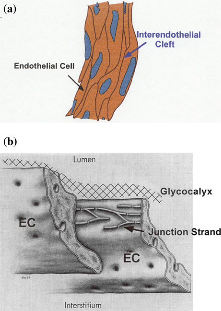
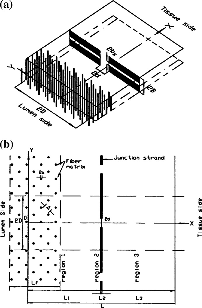
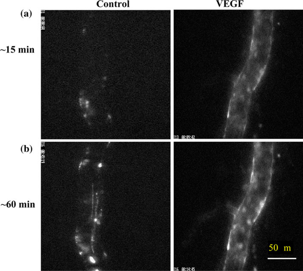
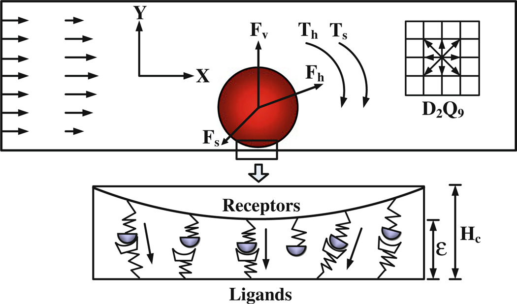
One critical step in tumor metastasis is tumor cell adhesion to the endothelium forming the microvessel wall. Understanding this step may lead to new therapeutic concepts for tumor metastasis. Vascular endothelium forming the microvessel wall and the glycocalyx layer at its surface are the principal barriers to, and regulators of the material exchange between circulating blood and body tissues. The cleft between adjacent ECs (interendothelial cleft) is the principal pathway for water and solutes transport through the microvessel wall in health. It is also suggested to be the pathway for high molecular weight plasma proteins, leukocytes and tumor cells across microvessel walls in disease. Thus the first part of the review introduced the mathematical models for water and solutes transport through the interendothelial cleft. These models, combined with the experimental results from in vivo animal studies and electron microscopic observations, are used to evaluate the role of the endothelial surface glycocalyx, the junction strand geometry in the interendothelial cleft, and the surrounding extracellular matrix and tissue cells, as the determinants of microvascular transport. The second part of the review demonstrated how the microvascular permeability, hydrodynamic factors, microvascular geometry and cell adhesion molecules affect tumor cell adhesion in the microcirculation.
Figures
Similar articles
-
Tumor Metastasis in the Microcirculation.Adv Exp Med Biol. 2018;1097:201-218. doi: 10.1007/978-3-319-96445-4_11. Adv Exp Med Biol. 2018. PMID: 30315547 Review.
-
Adhesion of malignant mammary tumor cells MDA-MB-231 to microvessel wall increases microvascular permeability via degradation of endothelial surface glycocalyx.J Appl Physiol (1985). 2012 Oct;113(7):1141-53. doi: 10.1152/japplphysiol.00479.2012. Epub 2012 Aug 2. J Appl Physiol (1985). 2012. PMID: 22858626 Free PMC article.
-
An electrodiffusion model for effects of surface glycocalyx layer on microvessel permeability.Am J Physiol Heart Circ Physiol. 2003 Apr;284(4):H1240-50. doi: 10.1152/ajpheart.00467.2002. Epub 2002 Dec 12. Am J Physiol Heart Circ Physiol. 2003. PMID: 12531731
-
A model for the modulation of microvessel permeability by junction strands.J Biomech Eng. 2003 Oct;125(5):620-7. doi: 10.1115/1.1611514. J Biomech Eng. 2003. PMID: 14618921
-
Microvascular solute and water transport.Microcirculation. 2005 Jan-Feb;12(1):17-31. doi: 10.1080/10739680590894993. Microcirculation. 2005. PMID: 15804971 Review.
Cited by
-
Comprehensive analysis of the whole coding and non-coding RNA transcriptome expression profiles and construction of the circRNA-lncRNA co-regulated ceRNA network in laryngeal squamous cell carcinoma.Funct Integr Genomics. 2019 Jan;19(1):109-121. doi: 10.1007/s10142-018-0631-y. Epub 2018 Aug 21. Funct Integr Genomics. 2019. PMID: 30128795
-
The mechanical responses of advecting cells in confined flow.Biomicrofluidics. 2020 May 4;14(3):031501. doi: 10.1063/5.0005154. eCollection 2020 May. Biomicrofluidics. 2020. PMID: 32454924 Free PMC article. Review.
References
-
- Akinaga T, Sugihara-Seki M, Itano T. Electrical charge effect on osmotic flow through pores. J. Phys. Soc. Jpn. 2008;77:053401.
-
- Antonetti DA, Wolpert EB, DeMaio L, Harhaj NS, Scaduto RC., Jr Hydrocortisone decreases retinal endothelial cell water and solute flux coincident with increased content and decreased phosphorylation of occludin. J. Neurochem. 2002;80:667–677. - PubMed
Publication types
MeSH terms
Grants and funding
LinkOut - more resources
Full Text Sources
