Review
. 2012 Jun;14(3):201-10.
doi: 10.1007/s11883-012-0244-1.

Atherosclerosis, inflammation, genetics, and stem cells: 2012 update

Affiliations
Review

Atherosclerosis, inflammation, genetics, and stem cells: 2012 update

Pascal J Goldschmidt-Clermont et al. Curr Atheroscler Rep. 2012 Jun.

Abstract

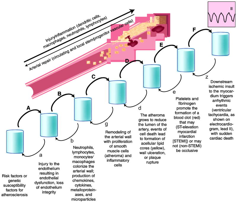
Atherosclerosis is a peculiar form of inflammation triggered by cholesterol-rich lipoproteins and other noxious factors such as cigarette smoke, diabetes mellitus, and hypertension. Genetics also play an important role in the disease, accounting for about 40% of the risk. Of surprise in recent years of post-human genome sequencing, atherosclerosis-relevant genes discovered by non-biased techniques (ie, genome-wide association studies), did not rehash previously suspected pathways of lipid metabolism, diabetes, or hypertension. Instead these studies highlighted genes relevant to mechanisms of inflammation and stem cell biology. Only a minority of implicated genes were linked to lipid and other cardiac risk factor genes. Although such findings do not contradict the fact that atherosclerosis is triggered and exacerbated by elevated lipids, atherosclerosis "new genes" suggest that the mechanism responsible for the development of arterial lesions is more complex than a simple response to injury, where injury is necessary, but perhaps not sufficient, for disease progression.


Figures

Figure 1
Figure 1
Effect of age on 10-year risk for coronary heart disease event. Ten-year risk as a function of age (in years) was calculated for an individual (man: blue; woman: red) who is a non-smoker, with no family history, no prior cardiac event, no diabetes mellitus, a fasting blood sugar of less than 100 mg/dL, a height of 5’ 8’’, a weight of 160 pounds, a waist circumference of less than 40 inches (man) or less than 35 inches (woman), a blood pressure of 120/70 mm Hg, and a cholesterol of 170 mg/dL (LDL 80 mg/dL, HDL 45 mg/dL, and triglycerides < 150 mg/dL). The risk increases by one to two orders of magnitude from 25 to 75 years of age in this individual with a relatively benign risk profile that was kept strictly unchanged over time. (Calculations made using the Heart Attack Risk Calculator from the American Heart Association. Available at https://www.heart.org/gglRisk/locale/en_US/index.html?gtype=health.)
Figure 2
Figure 2
Progression of atherosclerosis inflammation. Atherosclerosis as a complex disease process that can be deconstructed into a series of discrete events whose individual probability (A/α, B/β, C/γ, etc.) and timing vary with risk factors and intrinsic ability of self-repair for the artery. In general, the disease progression is presented as irreversible and one-directional as a function of time. However, for each step a reverse probability, even if rather small, does exist. Indeed, some individuals are genetically provided with a substantial ability for self-repair of their arteries, and consequently, even in the presence of potent risk factors, they remain sheltered from the consequences of atherosclerosis and related thromboembolic events.
Figure 3
Figure 3
Impact of effective arterial repair, or lack thereof, on arterial inflammation. A, In young individuals, the repair capacity of the arterial wall is almost always intact, with local and systemic reservoirs of stem/progenitor cells that are competent to repair an injured artery. As a consequence, an arterial injury triggers a limited inflammatory response, which is mediated by chemokines, cytokines, interleukins, and growth factors (such as vascular endothelial growth factor). The cellular inflammation response (cytoplasmic inflammasome) serves as a signal for the recruitment of inflammatory cells, but also stem/progenitor cells capable of repair, to the area of damaged arteries. Consequently, the artery is successfully repaired and the inflammation ceases (ie, a negative feedback loop). B, In older individuals, the repair capacity may become progressively exhausted. Once the bone marrow and other local reservoirs for repair-competent stem/progenitor cells are depleted, injury to the arterial wall triggers an inflammatory reaction that does not result in successful repair and, consequently, in the absence of negative feedback loop, inflammation continues unregulated (systemic elevation of C-reactive protein and other markers of inflammation). An unregulated inflammatory reaction at the level of the arterial wall contributes to larger atheroma, and unstable lesions ensue when the cellular inflammation response leads to rapid cell death (cytoplasmic apoptosome). In this situation, a positive feedback loop occurs, and unstable clinical events can occur as a consequence of plaque ulceration/rupture and thrombotic complications.
Figure 4
Figure 4
Effective arterial repair supported by periodic injections of bone marrow cells. Top panel: Enhanced culture step and bone marrow cells (BMC) repair efficacy. A, Proportion of total aorta containing atherosclerosis following injections of PBS, freshly harvested Lin BMCs, or enhanced culture (ECX) cells. B, Composite images for all analyzed aortas in each group; color indicates probability for the region to contain atherosclerosis (see color bar). From Song et al.[14]; with permission. Bottom panel: Cell biology or arterial repair. A, Cultured bone marrow (BM) cells from ROSA26 mice (expressing β-galactosidase in their nucleus) were injected intravenously into apolipoprotein E (ApoE)-deficient (knockout) C57 black 6 mice fed a high-fat diet. Shown are the aortae of recipient animals, after staining for β-galactosidase (blue coloration of positive cells). Blue cells have anchored in areas of the recipient aorta that otherwise would develop atherosclerosis. B, ApoE-knockout mice that did not receive ROSA26 cells (medium alone). In this case, no blue cells could be detected in the recipient aorta. C, Wild-type (WT) C57 black 6 mice who received cultured BMCs from ROSA26 mice do not display blue cells, indicating that attachment and homing of progenitor cells require an injury to the aorta (for ApoE knockout animals, injury corresponds to high circulating lipid levels). D, Oil Red O staining of ApoE knockout mice. Aorta from a mouse that received repeated injections of cultured BMCs showing reduced atherosclerosis (Oil Red O-positive lesions) of treated animals. E, Same staining as in D, for a mock-treated ApoE knockout mouse. F, H Fate of injected cultured BMCs (blue cells) in recipient aorta. G, I, A majority of the cells with a blue nucleus double-stain positive for markers of endothelial cells, such as PECAM (CD 31), indicating that most blue cells become endothelial cells that are incorporated within the surface area of the artery. Reproduced from Raucher et al. [13]

Similar articles

Cited by

References

    1. Brown MS, Goldstein JL. Koch's postulates for cholesterol. Cell. 1992;71(2):187–188. - PubMed
    1. Libby P, Ridker PM, Hansson GK. Progress and challenges in translating the biology of atherosclerosis. Nature. 2011;473(7347):317–25. Review. • This is a thorough review of the most recent information on atherosclerosis inflammation and its cellular and molecular agents. Novel clinical trials with anti-inflammatory drugs are also surveyed. - PubMed
    1. Karra R, Vemullapalli S, Dong C, et al. Molecular evidence for arterial repair in atherosclerosis. Proc Natl Acad Sci USA. 2005;102(46):16789–16794. - PMC - PubMed
    1. Ridker PM, Rifai N, Rose L, Buring JE, Cook NR. Comparison of C-reactive protein and low-density lipoprotein cholesterol levels in the prediction of first cardiovascular events. N Engl J Med. 2002;347(20):1557–1565. - PubMed
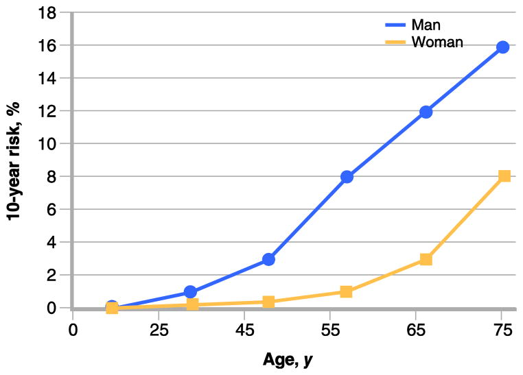
    1. American Heart Association. [Accessed December 2011.];Heart Attack Risk Calculator. Available at: https://www.heart.org/gglRisk/locale/en_US/index.html?gtype=health.

Publication types