. 2012 Sep;15(3):391-407.
doi: 10.1007/s10456-012-9268-y. Epub 2012 Apr 3.

Semaphorin 4D cooperates with VEGF to promote angiogenesis and tumor progression

Affiliations

Semaphorin 4D cooperates with VEGF to promote angiogenesis and tumor progression

Hua Zhou et al. Angiogenesis. 2012 Sep.

Retraction in

Abstract

The semaphorins and plexins comprise a family of cysteine-rich proteins implicated in control of nerve growth and development and regulation of the immune response. Our group and others have found that Semaphorin 4D (SEMA4D) and its receptor, Plexin-B1, play an important role in tumor-induced angiogenesis, with some neoplasms producing SEMA4D in a manner analogous to vascular endothelial growth factor (VEGF) in order to attract Plexin-B1-expressing endothelial cells into the tumor for the purpose of promoting growth and vascularity. While anti-VEGF strategies have been the focus of most angiogenesis inhibition research, such treatment can lead to upregulation of pro-angiogenic factors that can compensate for the loss of VEGF, eventually leading to failure of therapy. Here, we demonstrate that SEMA4D cooperates with VEGF to promote angiogenesis in malignancies and can perform the same function in a setting of VEGF blockade. We also show the potential value of inhibiting SEMA4D/Plexin-B1 signaling as a complementary mechanism to anti-VEGF treatment, particularly in VEGF inhibitor-resistant tumors, suggesting that this may represent a novel treatment for some cancers.


Figures
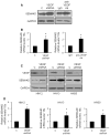
Fig. 1
Fig. 1
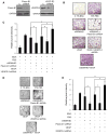
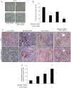
SEMA4D expression is elevated in HNSCC tumors and cell lines where VEGF function is inhibited by VEGF shRNA or anti-VEGF antibody. a Immunoblots for SEMA4D (upper panels) from tissue extracts of HN12 tumors comprised of control-infected cells (C) or cells infected with lentiviruses-expressing VEGF shRNA (VEGF shRNA, left panels) or from tumors where mice were treated with control antibody (IgG) or anti-VEGF antibody (anti-VEGF Ab) injections (right panels). b RNA was extracted from tumors from (a) to detect steady-state SEMA4D transcripts in a quantitative PCR analysis. The bar graph represents the ratio of SEMA4D mRNA to 18S mRNA, relative to controls, from three independent experiments (*p <0.05). c Immunoblot for VEGF (upper panel) and SEMA4D (middle panel) from HN12, 13 and 30 cells growing in culture, infected with control lentivirus (C) or lentivirus-expressing VEGF shRNA (VEGF shRNA). For all immunoblots, GAPDH was used as the loading control (lower panels). d RNA was extracted to detect steady-state SEMA4D transcripts in a quantitative PCR analysis from HN12, 13, and 30 cells from (c). The bar graphs represent the ratio of SEMA4D mRNA to 18S mRNA, relative to controls, from three independent experiments (*p <0.05)
Fig. 2
Fig. 2
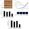
SEMA4D and VEGF act together to promote endothelial cell migration and tube formation in vitro. a Immunoblot analysis for the SEMA4D receptor Plexin-B1 (left, upper panel) and the VEGF receptor VEGFR-2 (right, upper panel) in lysates from HUVEC infected with empty vector control lentivirus (C) or virus coding for the appropriate shRNA. GAPDH was used as a loading control (lower panels). b Control or shRNA-expressing lentivirus-infected HUVEC from (a) were examined in a Boyden chamber for migration toward 0.1 % BSA (negative control), 10 % FBS (positive control), SEMA4D, VEGF, or both. c The results of the migration assay, expressed as pixel intensity of scanned stained migration membranes relative to negative controls. Error bars represent the standard deviation from three independent experiments (*p <0.05). d Control or shRNA-expressing lentivirus-infected HUVEC from (a) were plated on reconstituted basement membrane material in serum-free media with 0.1 % BSA (negative control), 10 % FBS (positive control), SEMA4D, VEGF or both and examined for formation of capillary tubes. e Quantification of the results of the tubulogenesis assay, measuring, and summing the length of all tubular structures observed in 10 random fields. Error bars represent the standard deviation from three independent experiments (*p <0.05)
Fig. 3
Fig. 3
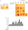
SEMA4D is a potent pro-angiogenic compound that cooperates with VEGF to enhance angiogenesis in vivo. a A DIVAA assay was performed in mice receiving injections of the indicated antibodies, measuring blood vessel growth into angioreactors containing reconstituted basement membrane material alone, or material mixed with VEGF, SEMA4D, or both. Negative and positive controls contain PBS and FGF, respectively (left panel). Photographs of representative angioreactors from the experiment are shown (right panels). b Quantification of blood vessel growth, as measured by FITC-lectin fluorescence (in arbitrary units, AU) from endothelial cell contents of each reactor, relative to negative controls (Y-axis). Error bars represent the standard deviation from four reactors (*p <0.05; **p <0.01)
Fig. 4
Fig. 4
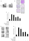
SEMA4D and VEGF are produced by HNSCC and promote endothelial cell migration and tube formation in vitro. a Immunoblot analysis for SEMA4D (upper panel, left) and VEGF (upper right) in lysates from HN12 cells infected with empty vector control lentivirus (C) or virus coding for SEMA4D shRNA or VEGF shRNA, where indicated. GAPDH was used as a loading control (lower panels). b Boyden chamber migration assay in HUVEC toward 0.1 % BSA (negative control), 10 % FBS (positive control) or serum-free media conditioned by HN12 cells, control-infected or infected with lenti-virus coding for SEMA4D shRNA, VEGF shRNA, or both. c Results of the migration assay from (b), quantified as the pixel intensity of scanned migration assay membranes relative to the negative control. Error bars represent the standard deviation from the averages from three wells (*p <0.05; **p <0.01). d HUVEC were plated on reconstituted basement membrane material in media containing 0.1 % BSA (negative control), 10 % FBS (positive control) or serum-free media conditioned by HN12 cells, control-infected or infected with lentivirus coding for SEMA4D shRNA, VEGF shRNA, or both, and examined for formation of capillary tubes. Representative photographs are shown. e Quantification of the results of the tubulogenesis assay, measuring, and summing the length of all tubular structures observed in 10 random fields. Error bars represent the standard deviation from three independent experiments (*p <0.05; **p <0.01)
Fig. 5
Fig. 5
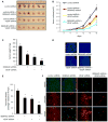
Production of SEMA4D by HNSCC cooperates with VEGF to promote tumor-induced angiogenesis. a HN12 cells infected with lentiviruses coding for control shRNA, SEMA4D shRNA, VEGF shRNA, or both were injected subcutaneously into nude mice along with a bolus of basement membrane extract. Representative tumors are shown at the time of kill. b The results of tumor volume measurement in cm3 are shown (n = 10; *p <0.05). c The results of tumor weights in mg are shown (n = 10; *, p <0.05; **p <0.01). d Immunofluorescence for CD31 as a measure of vascular density from tumors derived from HN12 cells infected with a control lentivirus (control shRNA), lentivirus-expressing SEMA4D shRNA or VEGF shRNA, or both. Nuclei are stained with DAPI. e Results of measurement of vascular content from these tumors, determined by the average number of vessels in 10 high-power fields (hpf) of CD31-stained sections, are quantified in the bar graph (*p <0.05; **p <0.01). f Immunofluorescence for vessels (green, top row) and pericytes (red, middle row) in the same tumor populations, looking for association of pericytes with endothelial cells (merge, bottom row) as an indicator of vessel integrity and maturity. Tumors from cells-expressing SEMA4D shRNA exhibit reduced numbers of vessels that fail to associate with pericytes. Tumors comprised of HN12 cells infected with VEGF shRNA-expressing lentivirus also fail to induce robust blood vessel growth, but those vessels are closely associated with a pericyte sheath
Fig. 6
Fig. 6
Production of SEMA4D and VEGF by HNSCC contributes to tumor cell proliferation and endothelial cell survival in tumor vasculature. a Immunohistochemistry for Ki-67, to measure proliferation of cells from tumors comprised of HN12 cells infected with control lentivirus (control shRNA), lentivirus-expressing SEMA4D shRNA, VEGF shRNA, or both. b Results of Ki-67 staining, expressed as percentage of positive cells observed from 10 high power fields. Error bars represent the standard deviation of the averages from three independent experiments (*p <0.05; **p <0.01). c The presence of cleaved caspase 3 (bottom row) was evaluated in tumors comprised of HN12 cells-expressing control shRNA, SEMA4D shRNA, VEGF shRNA or both, correlated with tumor sections stained for endothelial cells (CD31, top row). d Results of vascular apoptosis, expressed as percentage of vessels exhibiting cleaved caspase 3 observed from 10 high power fields, is shown in the bar graph (lower panel; p <0.05; **p <0.01)
Fig. 7
Fig. 7
Anti-SEMA4D therapy reduces growth and vascularity of tumors resistant to anti-VEGF treatment. a CT26 cells were injected subcutaneously into nude mice along with a bolus of basement membrane extract. Control mice were treated with IgG isotype control (IgG) or anti-VEGF antibody (anti-VEGF) for 18 days. Treatment for some mice receiving anti-VEGF antibody was changed to IgG (anti-VEGF + IgG) or anti-SEMA4D antibody (anti-VEGF + anti-SEM A4D) starting at day 9, extending until the end of the experiment (day 18). Representative tumors are shown at the time of kill. b The results of tumor volume measurement in cm3 are shown (n = 10; *p <0.05; **p <0.01). c The results of tumor weights in mg in mice treated with IgG or anti-VEGF antibody for the duration of the experiment or in mice treated with anti-VEGF antibody from days 1 to 9 and then switched to IgG or anti-SEMA4D antibody injections for days 9–18 (n = 10; *p <0.05). d Immunofluorescence for CD31 as a measure of vascular density from tumors derived from CT26 cells. e Results of measurement of vascular content from these tumors determined by the average number of vessels in 10 high-power fields (hpf) for CD31-stained sections (*p <0.05)

Similar articles

Cited by

References

    1. Zachary I. Vascular endothelial growth factor and anti-angiogenic peptides as therapeutic and investigational molecules. IDrugs. 2003;6:224–231. - PubMed
    1. Hiratsuka S, Minowa O, Kuno J, Noda T, Shibuya M. Flt-1 lacking the tyrosine kinase domain is sufficient for normal development and angiogenesis in mice. Proc Natl Acad Sci USA. 1998;95:9349–9354. - PMC - PubMed
    1. Lyden D, Hattori K, Dias S, Costa C, Blaikie P, Butros L, Chadburn A, Heissig B, Marks W, Witte L, Wu Y, Hicklin D, Zhu Z, Hackett NR, Crystal RG, Moore MA, Hajjar KA, Manova K, Benezra R, Rafii S. Impaired recruitment of bone-marrow-derived endothelial and hematopoietic precursor cells blocks tumor angiogenesis and growth. Nat Med. 2001;7:1194–1201. - PubMed
    1. Semenza GL. Regulation of hypoxia-induced angiogenesis: a chaperone escorts VEGF to the dance. J Clin Invest. 2001;108:39–40. - PMC - PubMed
    1. Fischer C, Jonckx B, Mazzone M, Zacchigna S, Loges S, Pattarini L, Chorianopoulos E, Liesenborghs L, Koch M, De Mol M, Autiero M, Wyns S, Plaisance S, Moons L, van Rooijen N, Giacca M, Stassen JM, Dewerchin M, Collen D, Carmeliet P. Anti-PlGF inhibits growth of VEGF(R)-inhibitor-resistant tumors without affecting healthy vessels. Cell. 2007;131:463–475. - PubMed

Publication types

LinkOut - more resources