

A new role for the P2X7 receptor: a scavenger receptor for bacteria and apoptotic cells in the absence of serum and extracellular ATP
- PMID: 22476940
- PMCID: PMC3360097
- DOI: 10.1007/s11302-012-9308-5
A new role for the P2X7 receptor: a scavenger receptor for bacteria and apoptotic cells in the absence of serum and extracellular ATP
Abstract
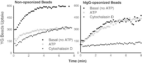
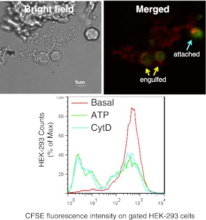
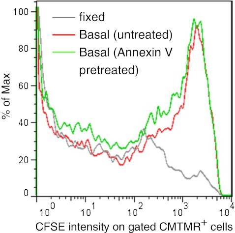
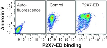
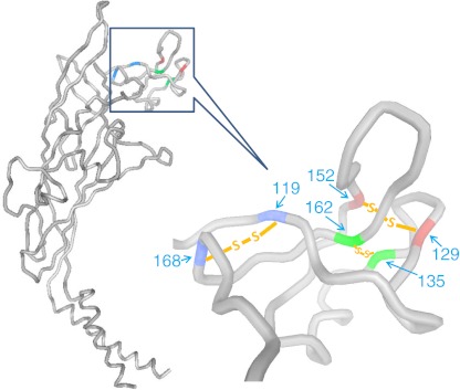
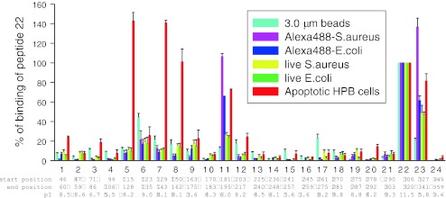
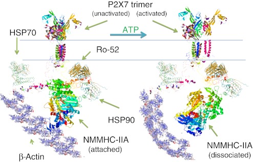
The P2X7 receptor is widely recognized to mediate the proinflammatory effects of extracellular ATP. However this receptor in the absence of ATP may have a function unrelated to inflammation. Our data show that P2X7 expressed on the surface of monocyte/macrophages or on epithelial HEK-293 cells greatly augments the engulfment of latex beads and live and heat-killed bacteria by effector phagocyte in the absence of ATP and serum. The expression of P2X7 on the effector also confers the ability to phagocytose apoptotic target cells and an accumulation of P2X7 can be seen at the attachment point to the target. Activation of the P2X7 receptor by ATP causes a slow dissociation (over 10-15 min) of nonmuscle myosin from the P2X7 membrane complex and abolishes further P2X7-mediated phagocytosis of these targets. The recent crystal structure of the homologous zebrafish P2X4 receptor shows an exposed "nose" of the ectodomain (residues 115-162) which contains three of the five disulfide bonds conserved in all P2X receptors. Three short biotin-labeled peptides mimicking sequence of this exposed region bound to apoptotic target cells but not to either viable cells or to other target particles. All three peptides contained one or two cysteine residues and their replacement by alanine abolished peptide binding. These data implicate thiol-disulfide exchange reactions in the initial tethering of apoptotic cells to macrophage and establish P2X7 as one of the scavenger receptors involved in the recognition and removal of apoptotic cells in the absence of extracellular ATP and serum.
Figures
Similar articles
-
P2X(7) is a scavenger receptor for apoptotic cells in the absence of its ligand, extracellular ATP.J Immunol. 2011 Sep 1;187(5):2365-75. doi: 10.4049/jimmunol.1101178. Epub 2011 Aug 5. J Immunol. 2011. PMID: 21821797
-
The P2X7-nonmuscle myosin membrane complex regulates phagocytosis of nonopsonized particles and bacteria by a pathway attenuated by extracellular ATP.Blood. 2010 Feb 25;115(8):1621-31. doi: 10.1182/blood-2009-11-251744. Epub 2009 Dec 9. Blood. 2010. PMID: 20007545
-
P2X7 from j774 murine macrophages acts as a scavenger receptor for bacteria but not yeast.Biochem Biophys Res Commun. 2016 Dec 2;481(1-2):19-24. doi: 10.1016/j.bbrc.2016.11.027. Epub 2016 Nov 8. Biochem Biophys Res Commun. 2016. PMID: 27833023
-
P2X7 as a scavenger receptor for innate phagocytosis in the brain.Br J Pharmacol. 2018 Nov;175(22):4195-4208. doi: 10.1111/bph.14470. Epub 2018 Oct 5. Br J Pharmacol. 2018. PMID: 30098011 Free PMC article. Review.
-
The human P2X7 receptor and its role in innate immunity.Tissue Antigens. 2011 Nov;78(5):321-32. doi: 10.1111/j.1399-0039.2011.01780.x. Tissue Antigens. 2011. PMID: 21988719 Review.
Cited by
-
P2X7 receptor modulates inflammatory and functional pulmonary changes induced by silica.PLoS One. 2014 Oct 13;9(10):e110185. doi: 10.1371/journal.pone.0110185. eCollection 2014. PLoS One. 2014. PMID: 25310682 Free PMC article.
-
Purinergic signaling in schistosomal infection.Biomed J. 2016 Oct;39(5):316-325. doi: 10.1016/j.bj.2016.06.006. Epub 2016 Nov 3. Biomed J. 2016. PMID: 27884378 Free PMC article. Review.
-
Possible protective role of the 489C>T P2X7R polymorphism in Alzheimer's disease.Exp Gerontol. 2014 Dec;60:117-9. doi: 10.1016/j.exger.2014.10.009. Epub 2014 Oct 16. Exp Gerontol. 2014. PMID: 25456845 Free PMC article.
-
Dexmedetomidine Alleviates Acute Stress-Induced Acute Kidney Injury by Attenuating Inflammation and Oxidative Stress via Inhibiting the P2X7R/NF-κB/NLRP3 Pathway in Rats.Inflammation. 2025 Feb;48(1):412-425. doi: 10.1007/s10753-024-02065-8. Epub 2024 Jun 19. Inflammation. 2025. PMID: 38896231
-
Characterization of P2X7R and its function in the macrophages of ayu, Plecoglossus altivelis.PLoS One. 2013;8(2):e57505. doi: 10.1371/journal.pone.0057505. Epub 2013 Feb 21. PLoS One. 2013. PMID: 23437395 Free PMC article.
References
-
- Herbomel P, Thisse B, Thisse C. Ontogeny and behaviour of early macrophages in the zebrafish embryo. Development. 1999;126(17):3735–3745. - PubMed
Publication types
MeSH terms
Substances
LinkOut - more resources
Full Text Sources
Miscellaneous
